Copyright
©The Author(s) 2021.
World J Stem Cells. Aug 26, 2021; 13(8): 1049-1057
Published online Aug 26, 2021. doi: 10.4252/wjsc.v13.i8.1049
Published online Aug 26, 2021. doi: 10.4252/wjsc.v13.i8.1049
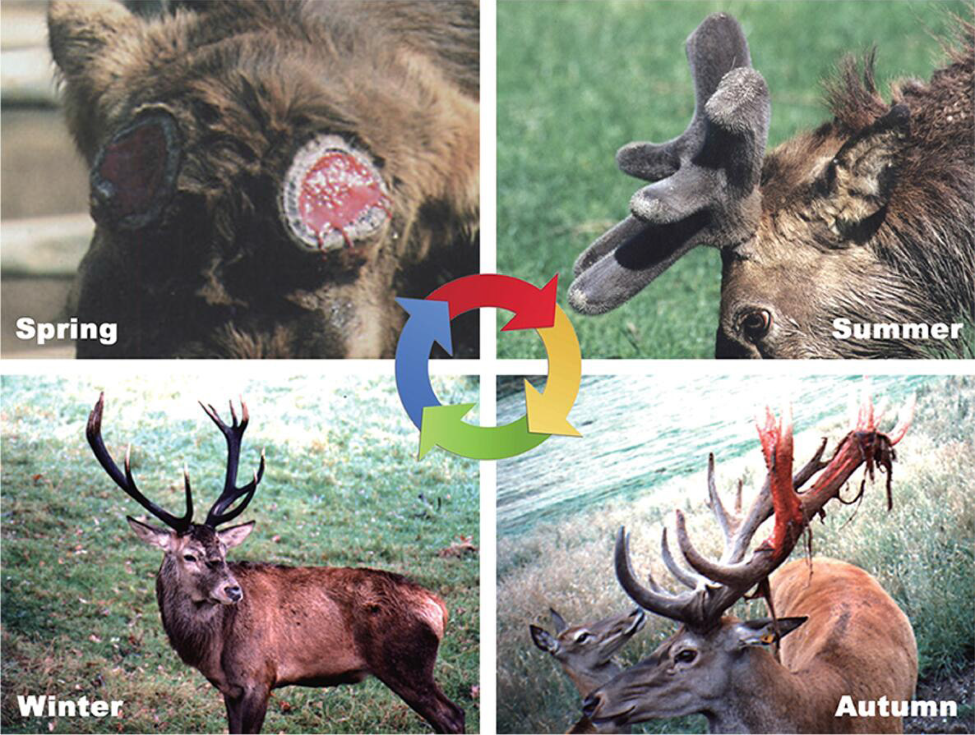
Figure 1 Antler regeneration cycle[11].
In spring, bony antlers drop off from their pedicle (permanent bony protuberance). Velvet antler regenerates immediately. In late spring and early summer, rapid antler growth occurs and antlers are covered with velvet skin in their growing phase. In autumn, antlers become completely calcified and the skin covering them starts to shed. In winter, dead bony antlers are attached to their living pedicles and eventually cast in spring next year, triggering a new round of antler regeneration. Citation: Li C, Chu W. The regenerating antler blastema: the derivative of stem cells resident in a pedicle stump. Front Biosci (Landmark Ed) 2016; 21: 455-467. Copyright © Frontiers in Bioscience. Published by Frontiers in Bioscience.
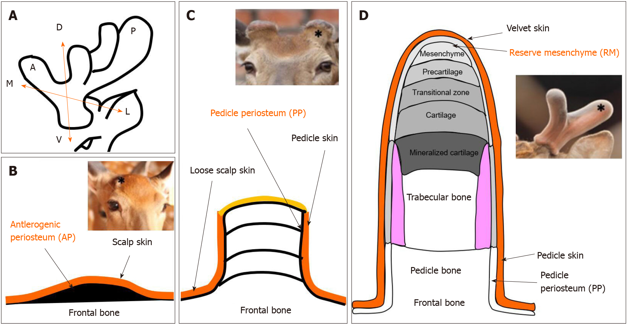
Figure 2 Schematic diagram of locations of antler stem cells.
A: Schematic diagram to show the three axes of the antler development: A ↔ P: Anterior-posterior axis; D ↔ V: Dorso-ventral axis; M ↔ L: Medio-lateral axis; B: The antlerogenic periosteum is present in the embryo and after birth as a localized thickening of the periosteum of the frontal bone; C: Regeneration of an antler initiated from the cells residing in the pedicle periosteum; D: Endochondral bone growth occurs at the distal tip, and cells in the reserve mesenchyme are responsible for rapid antler growth. Star in insert figure: Location of antler stem cells.
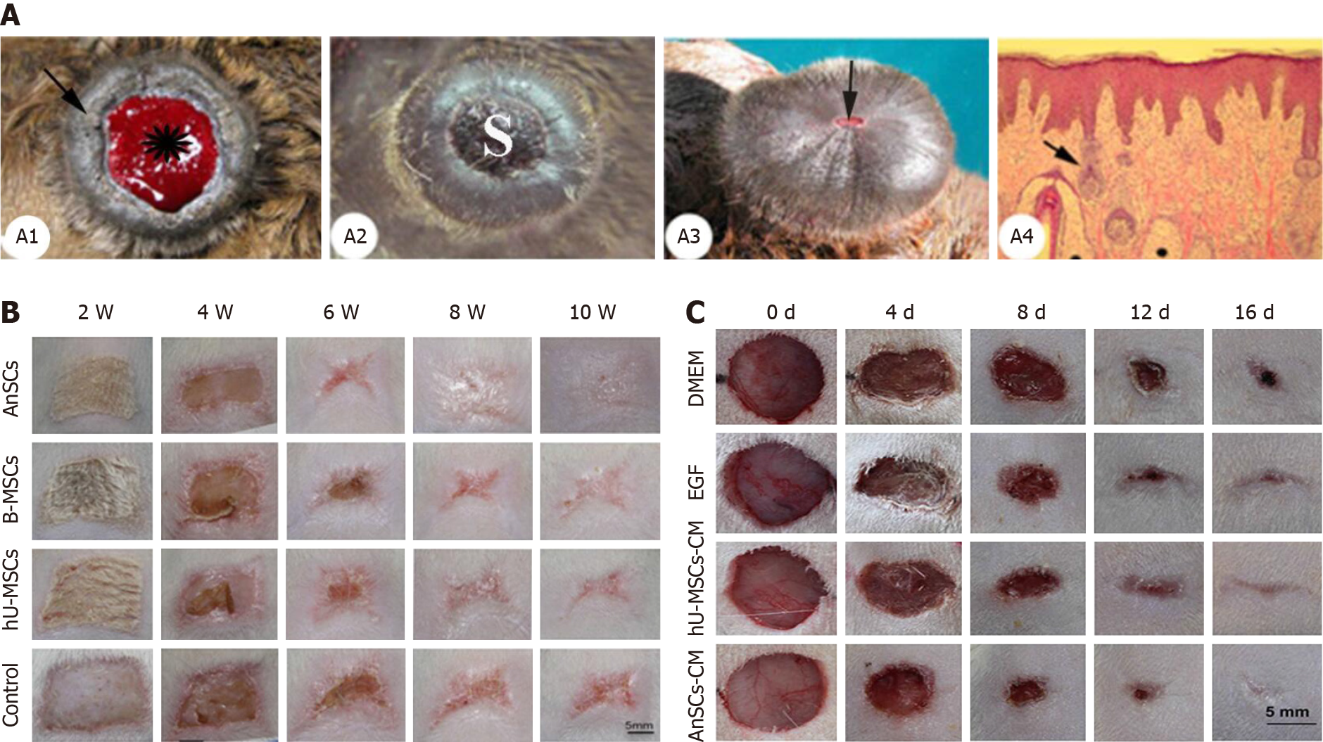
Figure 3 Antler stem cells-induced wound healing process in deer and rats[38-40].
A: Wound healing over the top of a pedicle stump following casting of a bony antler. A1: Pedicle with a fresh casting surface. A2: Apical surface of a pedicle a few days after hard antler casting. A3: Apical view of a late wound healing-stage pedicle. The scab becomes negligible. A4: Histological section of sagittal-cut healing skin; B and C: Gross morphological changes during wound healing occurring either via direct injection of antler stem cells (AnSCs) into the rats (B) or topical application of conditioned medium of AnSCs on to the wounds (C). hU-MCSs: Human mesenchymal stem cells; B-MSCs: Rat bone marrow mesenchymal stem cells; AnSCs: Antler stem cells. Citation for Figure 3A[38]: Li C, Suttie JM. Histological studies of pedicle skin formation and its transformation to antler velvet in red deer (Cervus elaphus). Anat Rec 2000; 260: 62-71. Copyright © Li Chunyi. Published by Mapsci Digital Publisher OPC Pvt Ltd. Citation for Figure 3B[39]: Rong X, Zhang G, Yang Y, Gao C, Chu W, Sun H, Wang Y, Li C. Transplanted Antler Stem Cells Stimulated Regenerative Healing of Radiation-induced Cutaneous Wounds in Rats. Cell Transplant 2020; 29: 963689720951549. Copyright © The author(s). Published by SAGE Publications Inc. Citation for Figure 3C[40]: Rong X, Chu W, Zhang H, Wang Y, Qi X, Zhang G, Wang Y, Li C. Antler stem cell-conditioned medium stimulates regenerative wound healing in rats. Stem Cell Res Ther 2019; 10: 326. Copyright © The author(s). Published by BioMed Central.
Figure 4 Regeneration of bone lesion after implantation of antler stem cells in mandibular bone lesions in rabbits[43].
Six and twelve months post antler stem cells implantation, restructuring of coarse fibrous bone into lamellar bone tissue occurred. Twenty-four months after implantation, mature lamellar bone developed, with visible osteons and two or three systemic lamellae around blood vessels. H&E staining in all sections. Citation: Cegielski M, Dziewiszek W, Zabel M, Dziegiel P, Kuryszko J, Izykowska I, Zatoński M, Bochnia M. Experimental xenoimplantation of antlerogenic cells into mandibular bone lesions in rabbits: two-year follow-up. In Vivo 2010; 24: 165-172. Copyright © International Institute of Anticancer Research. Published by International Institute of Anticancer Research.
- Citation: Zhang W, Ke CH, Guo HH, Xiao L. Antler stem cells and their potential in wound healing and bone regeneration. World J Stem Cells 2021; 13(8): 1049-1057
- URL: https://www.wjgnet.com/1948-0210/full/v13/i8/1049.htm
- DOI: https://dx.doi.org/10.4252/wjsc.v13.i8.1049


