修回日期: 2016-04-13
接受日期: 2016-04-20
在线出版日期: 2016-05-18
目的: 探讨血清铁调素(hepcidin, HEP)在急性胰腺炎(acute pancreatitis, AP)患者中的浓度变化和临床价值.
方法: 轻症急性胰腺炎(mild acute pancreatitis, MAP)104例、重症急性胰腺炎(severe acute pancreatitis, SAP)45例, 分别于入院时、入院后1、3、7 d用酶联免疫分析法检测HEP浓度, 以50例健康体检者为对照, 分析HEP与AP的临床相关性, 并绘制受试者工作特征曲线.
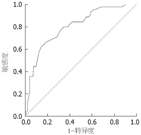
结果: AP患者入院时HEP浓度高于对照组(P<0.05); 入院后1、3、7 dSAP组HEP浓度高于MAP组(P<0.05); 入院时轻症、SAP组间HEP浓度无明显差异; HEP与血清C反应蛋白、APACHE-Ⅱ评分浓度正相关(P = 0.004、0.000), 与血钙浓度负相关(P = 0.003). 以入院后第3天APHEP浓度绘制的ROC曲线下面积为0.802, 以HEP浓度大于133 ng/mL为临界值, 以HEP诊断SAP的敏感度为95.6%, 特异度为61.5%.
结论: HEP与AP严重程度密切相关, 可能成为AP病情监测以及SAP早期识别的重要指标.
核心提示: 急性胰腺炎患者血清铁调素(hepcidin, HEP)浓度明显升高, 与APACHE-Ⅱ评分、C反应蛋白浓度正相关, 与钙离子浓度负相关. HEP浓度高于137 ng/mL提示重症急性胰腺炎.
引文著录: 沈阳, 薛成俊, 钟文贵, 陈志坚, 尤国莉, 薛勇. 血清铁调素在急性胰腺炎患者中的临床价值. 世界华人消化杂志 2016; 24(14): 2236-2240
Revised: April 13, 2016
Accepted: April 20, 2016
Published online: May 18, 2016
AIM: To investigate the changes of serum hepcidin in patients with acute pancreatitis and analyze its clinical significance.
METHODS: Blood samples from 104 patients with mild acute pancreatitis, 45 patients with severe acute pancreatitis and 50 healthy controls were collected, and serum hepcidin was measured by enzyme linked immunosorbent assay at admission, 1, 3, and 7 d after admission. The association of serum hepcidin with acute pancreatitis was analyzed. Receiver operating characteristic curve analysis was performed.
RESULTS: The median concentration of serum hepcidin in patients with acute pancreatitis at admission was significantly higher than that in healthy controls (P < 0.05). The differences in median concentrations of serum hepcidin between severe and mild acute pancreatitis were significant at 1, 3, and 7 d after admission (P < 0.05), but not at admission. Correlation analysis showed that serum hepcidin was positively correlated with C-reactive protein levels and APACHE-II score (P = 0.004, 0.000), but was negatively correlated with serum calcium levels (P = 0.003). The area under the ROC curve of serum hepcidin in patients with severe acute pancreatitis and mild acute pancreatitis at 3 d after admission was 0.802. With a cutoff value of 133 ng/mL, the overall sensitivity was 95.6%, and specificity was 61.5%.
CONCLUSION: Serum hepcidin correlates with the extent of pancreatitis. It can be used for monitoring the development of acute pancreatitis and may be a potential marker for early diagnosis of severe acute pancreatitis.
- Citation: Shen Y, Xue CJ, Zhong WG, Chen ZJ, You GL, Xue Y. Clinical significance of serum hepcidin in patients with acute pancreatitis. Shijie Huaren Xiaohua Zazhi 2016; 24(14): 2236-2240
- URL: https://www.wjgnet.com/1009-3079/full/v24/i14/2236.htm
- DOI: https://dx.doi.org/10.11569/wcjd.v24.i14.2236
近年来急性胰腺炎(acute pancreatitis, AP)发病率有不断升高趋势, 轻症急性胰腺炎(mild acute pancreatitis, MAP)绝大部分预后良好, 但重症急性胰腺炎(severe acute pancreatitis, SAP)起病急, 进展快, 总体病死率仍高达5%-10%[1]. 因此, AP的病情监测, 以及SAP的早期识别和判断, 对于指导治疗和改善预后具有举足轻重的作用. 至今AP严重程度的评估方法有很多, 比较经典的有Ranson、APACHE-Ⅱ和Balthazar CT等, 近年来有学者提出了BISAP、改良Marshall、MCTSI和C反应蛋白(C-reactive protein, CRP)等, 但这些指标要么较复杂, 要么有一定经济成本. 因此, 进一步寻找早期、准确、简单的AP严重度评价指标具有十分重要意义. Nemeth等[2]研究发现, 当机体受至炎症刺激时血清铁调素(hepcidin, HEP)浓度明显升高. 本研究通过动态监测AP患者血清HEP浓度的变化, 探讨HEP与AP临床相关性, 以及HEP在AP病情监测和严重度评估中的价值.
收集2014-09/2015-08南通大学附属建湖医院消化科AP患者149例, 女71例, 男78例, 年龄28-81岁, 中位年龄57.46岁±13.03岁. MAP患者104例, SAP患者45例. 另选择50例健康体检者作为对照组, 其中男29例、女21例, 中位年龄54.82岁±10.44岁, 均无明显不适主诉, 血常规、肝肾功能检查正常, 肝胆胰脾肾B超及胸部X线均正常. 所有AP病例与健康对照组间、SAP组与MAP组间年龄、性别构成均具有可比性. 纳入标准: 所有AP病例的诊断和分级均符合中国AP诊治指南; 所有病例的选取均为发病12 h内入院; 所有AP病例均为首次发病. 排除标准: 所有病例合并有与HEP浓度改变有关的疾病, 如缺铁性贫血、慢性胰腺炎、慢性肾功能不全、慢性肝病和自身免疫性疾病等; 服用糖皮质激素或免疫抑制剂的所有病例; 入院3 d内死亡或出院的所有病例; 不遵守研究协议内容及不能完成研究所需的检查测试的患者.
于患者入院时、1、3、7 d分别抽取外周静脉血, 3000 r/min, 离心10 min, 分离出血清后置-80 ℃冰箱保存. 酶联免疫试剂盒购于美国Bioswamp, 按说明书步骤操作, 运用竞争性同相酶联免疫分析法测定标本HEP浓度. 对健康体检者血标本采用以上相同的检测方法.
统计学处理 采用SPSS16.0统计分析软件, 计量资料两组间平均数的比较采用t检验; 构成比的比较采用χ2检验; 采用Pearson检验对AP患者血清HEP浓度与CRP、血清钙离子浓度及APACHE-Ⅱ评分行相关性分析, 并绘制受试者操作特性曲线(receiver operating characteristic curve, ROC曲线), 计算ROC曲线下面积(area under the ROC curve, AUC), P<0.05为差异有统计学意义.
入院时AP患者血淀粉酶、HEP水平较健康对照组明显升高, 差异具有统计学意义(P<0.05), 而年龄、性别、白蛋白和血红蛋白浓度与健康对照组比较差异无明显统计学意义(P>0.05)(表1).
| 分组 | AP组 | 健康对照组 | P值 |
| 年龄(岁) | 57.46±13.03 | 54.82±10.44 | 0.20 |
| 性别(男/女) | 71/78 | 29/21 | 0.33 |
| 淀粉酶(U/L) | 1309.00±1295.55 | 68.26±18.80 | 0.00 |
| 白蛋白(g/L) | 36.38±2.29 | 35.88±1.77 | 0.16 |
| 血红蛋白(g/L) | 139.98±15.04 | 137.22±8.69 | 0.12 |
| 入院时铁调素(ng/mL) | 115.88±15.56 | 50.26±13.92 | 0.00 |
入院后1、3、7 d SAP组血清HEP浓度分别为129.49 ng/mL±15.57 ng/mL、158.07 ng/mL±11.73 ng/mL、168.87 ng/mL±18.43 ng/mL, MAP组HEP浓度分别为124.58 ng/mL±12.60 ng/mL、142.62 ng/mL±15.10 ng/mL、132.62 ng/mL±14.52 ng/mL. 不同时间点两组间HEP浓度比较均具有统计学差异(P<0.05), 但入院时MAP与SAP两组间HEP浓度比较, 差异无统计学意义(表2).
| 分组 | MAP组 | SAP组 | P值 |
| 入院时 | 114.35±15.48 | 119.42±15.34 | 0.067 |
| 1 d | 124.58±12.60 | 129.49±15.57 | 0.044 |
| 3 d | 142.62±15.09 | 158.07±11.73 | 0.000 |
| 7 d | 132.62±14.52 | 168.87±18.43 | 0.000 |
AP患者HEP与CRP、APACHE-Ⅱ评分正相关(r = 0.233、0.369, P = 0.004、0.000), 与血钙浓度呈负相关(r = -0.246, P = 0.003).
AP患者入院后第3天HEP浓度绘制的ROC曲线下面积为0.802, 以HEP>137 ng/mL为临界值, 诊断SAP的敏感度为95.6%, 特异度为61.5%, 95%可信区间为0.726-0.878(图1).
HEP是一种小肽类激素, 主要由肝细胞合成, 前体是含有84个氨基酸组成的多肽, 后被信号肽酶剪接成无活性的前体, 再经激素原转化酶切割为含25个氨基酸的活性肽, 最后进入血液循环发挥作用, 并可随尿液排出体外[3,4]. 他是机体铁代谢重要的调节因子, 与体内铁呈负反馈关系[5-7]. Nicolas等[8]研究发现HEP除了与铁储存、贫血有关, 还受缺氧、炎症和细胞因子等多个因素直接或间断调控[8-10], 同时其本身也参与炎症反应过程. 有研究[11-13]发现在急性感染患者中HEP水平明显升高, 认为HEP是急性时相蛋白之一. 本研究通过动态监测AP患者HEP浓度, 探讨其与AP临床相关性, 以及其在病情监测和严重度评估中的作用, 而国内相关报道较少.
本研究发现入院时AP组HEP浓度高于健康对照组, 且SAP组于入院后1、3、7 d HEP浓度明显高于MAP组. 与Arabul等[14]研究结果基本一致. AP时HEP浓度升高的具体机制尚不明确, 可能系AP在发生发展过程中, 产生了大量的炎症介质, 如TNF、IL-6、IL-8和CRP等, 而这些炎症介质能够强烈刺激HEP的表达. 目前以IL-6的研究最为深入[15], 并认为IL-6介导炎症反应过程中的HEP升高的主要因子[11]. 研究[2]发现IL-6与首先与其受体结合, 其次激活JAKs, 进而将STAT蛋白磷酸化, 磷酸化的STAT蛋白3进入细胞核, 直接与HEP基因启动因子相应位点结合, 从而促进HEP的表达. 本研究还发现MAP患者HEP于入院后3 d达高峰, 而SAP患者HEP于本研究第7天达高峰, 与Arabul等[14]研究发现HEP高峰均处于第7天不完全一致. 分析其原因可能系由于MAP入院后于病程早期炎症介质大量释放, 但由于禁食、胃肠减压和药物使用等措施积极干预, 炎症反应被即时阻断. 而重症胰腺炎患者由于胰腺微循环障碍、肠屏障功能减退, 细菌易位等形成"第二次打击", 级联式"瀑布"反应未能及时终止, 大量炎性介质再次释放, 引起全身炎症反应综合征和器官功能衰竭发生[16], 从而导致SAP患者HEP浓度进行性升高, 并于本研究第7天达高峰.
APACHE-Ⅱ评分、CRP和血清钙离子浓度在临床评估AP严重程度方面虽有其局限性, 但均具有一定的应用价值[17,18]. 本研究发现, 血清HEP与APACHE-Ⅱ评分、CRP呈正相关, 与血清钙离子浓度呈负相关, 且入院后第3天HEP的ROC曲线下面积为0.802, 以HEP>137 ng/mL为临界值, 诊断SAP的敏感度为95.6%, 特异度为61.5%, 95%可信区间为0.726-0.878. 所以HEP可能成为AP病情监测以及SAP早期识别的重要指标. 由于本研究为单一医院数据, 研究对象有限, 所以统计把握度受限, 其具体调控机制也有待进一步深入研究, 临床应用尚需更加深入的临床试验的补充.
急性胰腺炎(acute pancreatitis, AP)发病率不断增加, 部分患者病情凶险, 进展迅速, 病死率仍高达5%-10%. 目前评估AP病情的指标存在复杂、价格昂贵等局限. 近年来发现血清铁调素(hepcidin, HEP)是评估AP严重程度的重要指标.
黄颖秋, 教授, 本溪钢铁(集团)总医院消化内科
AP患者HEP浓度明显升高, 目前研究较多的是白介素-6经由JAK/STAT通路调控其表达, 但确切机制待尚进一步深入研究.
Arabul等研究认为HEP浓度在AP患者中明显升高, 于病程第7天达至高峰, 是判断病情严重程度的重要指标, 而且优于C反应蛋白(C-reactive protein, CRP)的价值, 与本研究结果基本一致.
本文选择国内外认可的检测方法, 增加样本量, 同时结合CRP、APACHE-Ⅱ评分和钙离子浓度, 以及计算ROC曲线下面积(area under the ROC curve, AUC)进一步提高HEP诊断重症急性胰腺炎的临床可靠性.
本文通过探讨HEP在AP病情监测和严重度评估中的作用, 有助于早期、准确地评估AP患者的病情, 从而及时采取相应的干预措施, 改善患者预后.
ROC曲线: 即受试者工作特征曲线, 最初用于评价雷达性能, 又称为接收者操作特性曲线. ROC曲线是根据一系列不同的二分类方式(分界值或决定阈), 以真阳性率(灵敏度)为纵坐标, 假阳性率(1-特异度)为横坐标绘制的曲线.
本文初步探讨了HEP对AP严重程度的评估价值, 有一定临床意义.
编辑: 于明茜 电编:闫晋利
| 1. | 中华医学会消化病学分会胰腺疾病学组, 《中华胰腺病杂志》编辑委员会, 《中华消化杂志》编辑委员会. 中国急性胰腺炎诊治指南(2013年, 上海). 中华胰腺病杂志. 2013;13:73-78. |
| 2. | Nemeth E, Valore EV, Territo M, Schiller G, Lichtenstein A, Ganz T. Hepcidin, a putative mediator of anemia of inflammation, is a type II acute-phase protein. Blood. 2003;101:2461-2463. [PubMed] [DOI] |
| 3. | Park CH, Valore EV, Waring AJ, Ganz T. Hepcidin, a urinary antimicrobial peptide synthesized in the liver. J Biol Chem. 2001;276:7806-7810. [PubMed] [DOI] |
| 4. | Hunter HN, Fulton DB, Ganz T, Vogel HJ. The solution structure of human hepcidin, a peptide hormone with antimicrobial activity that is involved in iron uptake and hereditary hemochromatosis. J Biol Chem. 2002;277:37597-37603. [PubMed] [DOI] |
| 5. | Schmidt PJ. Regulation of Iron Metabolism by Hepcidin under Conditions of Inflammation. J Biol Chem. 2015;290:18975-18983. [PubMed] [DOI] |
| 6. | Krawiec P, Pac-Kozuchowska E. [The role of hepcidin in iron metabolism in inflammatory bowel diseases]. Postepy Hig Med Dosw (Online). 2014;68:936-943. [PubMed] |
| 8. | Nicolas G, Chauvet C, Viatte L, Danan JL, Bigard X, Devaux I, Beaumont C, Kahn A, Vaulont S. The gene encoding the iron regulatory peptide hepcidin is regulated by anemia, hypoxia, and inflammation. J Clin Invest. 2002;110:1037-1044. [PubMed] [DOI] |
| 9. | Singh B, Arora S, Agrawal P, Gupta SK. Hepcidin: a novel peptide hormone regulating iron metabolism. Clin Chim Acta. 2011;412:823-830. [PubMed] [DOI] |
| 10. | Petrova J, Manolov V, Vasilev V, Tzatchev K, Marinov B. Ischemic stroke, inflammation, iron overload - Connection to a hepcidin. Int J Stroke. 2016;11:NP16-NP17. [PubMed] [DOI] |
| 11. | Armitage AE, Eddowes LA, Gileadi U, Cole S, Spottiswoode N, Selvakumar TA, Ho LP, Townsend AR, Drakesmith H. Hepcidin regulation by innate immune and infectious stimuli. Blood. 2011;118:4129-4139. [PubMed] [DOI] |
| 12. | Atkinson SH, Armitage AE, Khandwala S, Mwangi TW, Uyoga S, Bejon PA, Williams TN, Prentice AM, Drakesmith H. Combinatorial effects of malaria season, iron deficiency, and inflammation determine plasma hepcidin concentration in African children. Blood. 2014;123:3221-3229. [PubMed] [DOI] |
| 13. | Bagu ET, Layoun A, Calvé A, Santos MM. Friend of GATA and GATA-6 modulate the transcriptional up-regulation of hepcidin in hepatocytes during inflammation. Biometals. 2013;26:1051-1065. [PubMed] [DOI] |
| 14. | Arabul M, Celik M, Aslan O, Torun S, Beyazit Y, Alper E, Kandemir A, Ünsal B. Hepcidin as a predictor of disease severity in acute pancreatitis: a single center prospective study. Hepatogastroenterology. 2013;60:595-600. [PubMed] [DOI] |
| 15. | Qian ZM, He X, Liang T, Wu KC, Yan YC, Lu LN, Yang G, Luo QQ, Yung WH, Ke Y. Lipopolysaccharides upregulate hepcidin in neuron via microglia and the IL-6/STAT3 signaling pathway. Mol Neurobiol. 2014;50:811-820. [PubMed] [DOI] |
| 16. | Lankisch PG, Bruns A, Doobe C, Weber-Dany B, Maisonneuve P, Lowenfels AB. The second attack of acute pancreatitis is not harmless. Pancreas. 2008;36:207-208. [PubMed] [DOI] |
| 17. | Zrnić IK, Milić S, Fisić E, Radić M, Stimac D. [C-reactive protein and lactate dehydrogenase as single prognostic factors of severity in acute pancreatitis]. Lijec Vjesn. 2007;129:1-4. [PubMed] |
| 18. | Yeung YP, Lam BY, Yip AW. APACHE system is better than Ranson system in the prediction of severity of acute pancreatitis. Hepatobiliary Pancreat Dis Int. 2006;5:294-299. [PubMed] |


