Basic Research Open Access
Copyright ©The Author(s) 2003. Published by Baishideng Publishing Group Inc. All rights reserved.
World J Gastroenterol. Jun 15, 2003; 9(6): 1282-1286
Published online Jun 15, 2003. doi: 10.3748/wjg.v9.i6.1282
Isolation and analysis of a novel gene over-expressed during liver regeneration
Yu-Chang Li, Cun-Shuan Xu, Wen-Qiang Li, College of Life Science, Henan Normal University, Xinxiang 453002, Henan Province, China
Wu-Lin Zhu, Xinxiang Medical College, Xinxiang 453002, Henan Province, China
Author contributions: All authors contributed equally to the work.
Supported by a grant from Key Scientific and Technical Problem of Henan Province No.0122031900
Correspondence to: Dr. Yu-Chang Li, College of Life Science, Henan Normal University, Xinxiang 453002, Henan Province, China. ycli_us@yahoo.com
Telephone: +86-373-3328084 Fax: +86-373-3326609
Received: December 5, 2002
Revised: March 1, 2003
Accepted: March 3, 2003
Published online: June 15, 2003

Abstract

AIM: To isolate and analyze a novel gene over-expressed during liver regeneration.

METHODS: Total RNA of regenerating liver was extracted from liver tissue after 0-4-36-36-36 hr short interval successive partial hepatectomy (SISPH). Reverse transcription-polymerase chain reaction was used to synthesize double strand cDNA, after the tissue was digested by proteinase K and Sfi A/B. The double-strand cDNA was ligated to λTriplEx2. λphage packaging reaction was performed and E. coli XL1-Blue was infected for titering and amplifying. One expressed sequence tag was probed by Dig and phage in situ hybridization was carried out to isolate positive clones. Positive recombinant λTriplEx2 was converted to the corresponding pTriplEx2, and bioinformatics was used to analyze full-length cDNA.

RESULTS: We isolated a novel full-length cDNA during liver regeneration following SISPH.

CONCLUSION: We have succeeded in cloning a novel gene, based on bioinformatics. We postulate that this gene may function in complicated network in liver regeneration. On the one hand, it may exert initiation of liver regeneration via regulating nitric oxide synthesis. On the other hand, it may protect damaged residue lobus following SISPH.


INTRODUCTION

Liver regeneration is a system suitable for investigating normally regulated growth[1-4]. After surgical removal of 70% of the mass of a healthy liver (two-thirds hepatectomy), residual tissue enlarges to make up for the mass of removed substance in entirety, in small animals, this process usually lasts 5 to 7 d[5-7].

The growth response after partial hepatectomy is governed by priming and progression through the cell cycle[2,6,8]. The priming phase coincides with loss of growth inhibition and represents the G0 to G1 transition, whereas the progression phase acts on promoting cell replication and represents the G1 to S transition. Priming involves the activation of a group of non-specific factors, which are necessary, but not sufficient for the S phase completion; they comprise the nuclear factor for kappa chains (NF-κ) in B cells, signal transducer and activator of transcription-3 (STAT3), activator protein-1 (AP-1), CCAAT enhancer binding protein, and several immediate early genes like epithelial growth factor (EGF), tumor necrosis factor-alpha (TNF-α, IL-6), insulin, and matrix changes[9-15]. The priming step is reversible until the cells have crossed the so-called G1 checkpoint, the cells thereupon being irreversibly committed for replication. Moreover, the initiation of the growth response depends on complex interactions among hepatocytes and nonparenchymal cells, the extracellular matrix (ECM), endocrine, autocrine, paracrine, and neuroregulatory factors, oxygen free radicals, metabolites, and nutrients[2,6,9,16]. Progression signals include hepatocyte growth factor (HGF), transforming growth factor-alpha (TGF-α), EGF and insulin. The regulation of hepatic regenerative process depends on a number of myriad factors that ultimately modulate immediate-early, delayed-early, and liver-specific gene expression[2,6,9,17-20].

The termination of hepatic regeneration still remains an enigma. A variety of factors have been touted as growth inhibitors/terminators during the regenerative response once recovery of the liver mass has been achieved.TGF-β and activins are regarded as potent inhibitors involved in the termination response[21,22]. TGF-β is a fibrogenic cytokine secreted by hepatic stellate cells, TGF-β mRNA, almost undetectable in normal liver, increases within 3-4 h after partial hepatectomy, and attains a plateau after 48-72 h. TGF-β1 overexpression in transgenic mice inhibits the abundance of the cyclin-dependent kinase activating tyrosine phosphatase cdc25A protein, and is associated with increased binding of histone deacetylase 1 to p130 in the liver[23]. Activin A (the homodimer of the inhibin βA chain) inhibits but follistatin (anactivin-binding protein) promotes hepatocyte proliferation. Whereas activin A is a negative regulator of hepatocyte proliferation, mice deficient in both activin βC and activin βE, are not different from wild-type mice with respect to liver development and the regenerative response after partial hepatectomy. The activin system also plays a significant role in the dynamics of the ECM, in particular fibronectin; ECM components are reconstructed during desinualization and hepatocyte cluster formation. After rat liver injury and partial hepatectomy, hepatocyte activin A receptors are down-regulated at 24 hr and normalized at 72 hr. This phenomenon may be involved in rendering hepatocytes responsive to mitogenic stimuli, whereas increased activin A levels stimulate stellate cell production of fibronectin, important for the growth and proper placement of regenerating hepatocytes[24].

Despite the research efforts, our knowledge on the regulatory mechanisms of cell growth, differentiation and tissue organization is limited. There may be some unknown factors that may play important roles in the process of liver regeneration. To acquire a better understanding on the mechanisms involved, some researchers have established a series of successive partial hepatectomy (SPH) models. These mainly include the long interval successive partial hepatectomy (LISPH) in which an interval of more than three weeks is applied as described by Wu et al, the SPH model where a one-third hepatectomy 2 wk after a two-thirds hepatectomy is performed as described by Takeshi et al[25], and the short interval successive partial hepatectomy (SISPH) model (0-4-40-76-112 hr) which has an interval of 4 and/or 36 h because the cells would re-enter into dedifferentiation stage after 4 hr and reach the peak of cell division after 36 hr following partial hepatectomy as described by Xu et al[26]. Studies have demonstrated that SISPH could provide more useful materials for analyzing the mechanism of liver regeneration[27]. We have begun identifying and characterizing some genes that are strongly expressed in liver regeneration. We took 0 h and 112 hr as driver and tester respectively in 0-4-36-36-36hr SISPH model and a suppression subtracted hybridization (SSH) method was performed[28-30]. Then we constructed a forward-subtractive cDNA library from which we have cloned 53 up-regulated expressed sequence tags (ESTs). Among these, nine ESTs were 100% homologous to GenBank and 44 ESTs were homologous to GenBank. one of these ESTs was found to be a novel gene in GenBank. In the present study, we have used one subtracted probe from suppression subtracted library in liver regeneration and isolated its full-length cDNA from cDNA library by the phage in situ hybridization method. Based on bioinformatics, we suggest that it might play important roles in the regulation of initiation of liver regeneration.

MATERIALS AND METHODS
Establishment of the SISPH model

Adult Spargue-Dawfey rats (weighing 200-250 g) were provided by the Experimental Animal House of Henan Normal University, and the 0-4-36-36-36 hr SISPH model was made according to the method described by Xu et al[26]. Lobus external sinister and lobus centralis sinister, lobus centralis, lobus dexter and lobus candatus were removed one by one at four different time points, i.e. at 4, 36, 36 and 36 hr (total time: 4 hr, 40 hr, 76 hr, 112 hr), respectively. The fourth resected liver lobus was washed with precooled phosphate- buffered saline (PBS) thoroughly, then the sample was frozen in liquid nitrogen and transferred to the -80 °C freezer for storage.

Primers

A1: 5'-ATTCTAGAGGCCGAGGCGGCCGACATG-d (T)30 N-3'

A2: 5'-AAGCAGTFFTATCAACGCAGAGT-3'

A3: 5'-TCGAGCGGCCGCCCGGGCAGGT-3'

A4: 5'-AGCGTGGTCGCGGCCGAGGT-3'

A5: 5'-TCCGAGATCTGGACGAGC-3'

A6: 5'-TAATACGACTCACTATAGGG-3'

Among these primers, A1 was used for synthesis of first cDNA strand, A1/A2 were used to amplify dscDNA, A3/A4 were used to probe expressed sequence tag with Dig, A5/A6 were used to detect full length cDNA.

RNA Isolation

Total RNA was isolated from 112 h liver tissue samples following SISPH by the method described by Chomczynski and Sacchi[31]. Tissues were homogenized and extracted twice with acidic guanidinium isothiocyanate-phenol-chloroform. The poly(A)+ RNA fraction was isolated by oligo-dT cellulose chromatography (Pharmacia Diagnostics AB, Uppsala, Sweden). The purity and integrity of total RNA were monitored by absorbance of ultraviolet spectrometer at 260/280 nm, and electrophoresis was carried out on a denaturing formaldehyde agarose gel and the gel was stained with ethidium bromide.

RT-PCR

300 ng mRNA was reversely transcripted to single-stranded cDNA by Powerscriptase at 42 °C for 1hr. First-strand cDNA was synthesized with a Sfi IB-oligo(dT) adapter-primer (A1). The resulting single strand cDNA was amplified by PCR using CDSIII/3'primer (A1) and 5'primer (A2) following parameters: 94 °C for 45 s, 68 °C for 6 min.

cDNA library construction

The double strand cDNA synthesis and library construction were carried out mainly according to the manual of SMART cDNA Library Construction Kit (Clontech, Heidelberg, Germany). After second-strand synthesis and ligation of Sfi IA adapters, cDNA was digested by Sfi IA/Sfi IB, generating cDNA flanked by Sfi IA sites at 5' ends and/Sfi IB sites at the 3' ends. Digested cDNAs were size-fractionated with Sephacryl S-500 spin columns and ligated into the λTriplEx2 express vector predigested by Sfi IA/Sfi IB. The resulting concatomers were packaged by using Gigapack Gold packaging extracts. After titration, aliquots of primary packaging mixture were stored in 7% DMSO at -80 °C as primary library stocks. At the same time, the ratio of white (recombinant) to total (white + blue (nonrecombinant)) assay was determined[32]. The remainder was amplified to establish stable library stocks.

Probe labeling and Phage in situ hybridization

A novel EST related to liver regeneration was probed with A3/A4 primers according to the user manual of Dig Probe Synthesis kit (Roche Diagnostics, Mannheim, Germany). Thermal cycle parameters were 94 °C for 2 min, 25 cycles including a denaturation step at 94 °C for 15sec, an annealing step at 68 °C for 30sec, extension at 72 °C for 1 min and 30sec, and a final extension step at 72 °C for 7 min. The amplified phage cDNA library was diluted in 1 × lamda dilution buffer to obtain a concentration of 104 pfu/mL and then to infect E. coli XL1-Blue. Host bacterial cells were absorbed by phage at 37 °C for 15 min and the indicated volume of melted LB top agarose/MgSO4 was added. The mixture was inverted once and poured onto a prewarmed, dry LB/MgSO4 plate. The plate was inverted and incubated at 37 °C until plaques became distinctly visible. A nylon filter was numbered and placed onto the LB soft top agarose, then the filter was marked in three asymmetric locations. After 2 min, the filter was peeled off carefully. The filter was placed in petri dishes orderly containing DNA denaturing solution for 5 min, neutralizing solution for 5 min, or 2 × SSC for 5 min. Hybridization and wash procedures were performed according to Sambrook[33]. When positive signal appeared the process was performed as above for the secondary and tertiary screening until single clone was obtained. Then recombinant λTriplEx2 was converted to the corresponding pTriplEx2. Plasmid was extracted and sent to TaKaRa (Dalian, China) for sequencing analysis.

RESULTS
RT-PCR

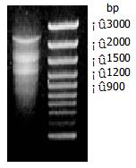
The mRNA was isolated from the 112 h liver tissue following SISPH. A modified oligo (dT) primer (A1 primer) primed the first-strand synthesis reaction. The resulting single strand cDNA was amplified by PCR using CDSIII/3'primer (A1) and 5'primer (A2) derived from SMART oligonucleotide (Figure 1).

Figure 1
Figure 1 Gel electrophoresis of double strand cDNA. Lane 1: Double strand cDNA; lane 2: DNA mass marker.
Determination of cDNA library

The unamplified library and amplified library were titered when E.coli XL1-Blue was infected by λphage. The plaques were counted and cDNA library was calculated respectively as follows: pfu/mL = number of plaques × dilution × 103μL/ml/μL of diluted phage plated. Results showed that umamplified cDNA library and amplified library were 6 × 10⁶ pfu/mL and 6 × 10¹⁰ pfu/mL respectively. X-gal/IPTG was used to assess recombination efficiency. Among 500-1000 plaques, the ratio of white (recombinant) to total (recombinant and blue (nonrecombinant)) was 99.52%. These data suggest that the cDNA library was successfully established.

Screening result and bioinformatics

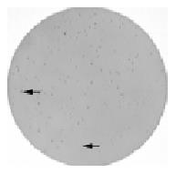
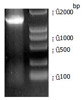
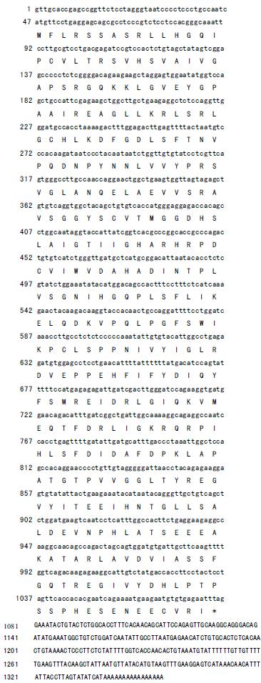
Figure 2 shows the tertiary positive clone screened by phage in situ hybridization. After conversion of a recombinant λTriplEx2 to the corresponding pTriplEx2, the tertiary positive clones were analyzed by PCR with the sequencing primers A5/A6.The length detected by electrophoresis was consistent with the sequencing result and its full length cDNA was 1354 bp (Figure 3). We identified that its ORF region encodes 344 amino acids, its start codon AUG was in 47-49 nucleotide and its termination codon TAG was in 1079-1081 nucleotides, and the sequence followed by TAG was polyA (Figure 4). It was compared with other sequences in the GenBank through Internet by BLAST and found to be 98.8% homologous to mouse arginase. The sequence has been deposited in the GenBank database (accession No. AF508019).

Figure 2
Figure 2 Tertiary screening result of positive clones. Arrows point the positive clones.
Figure 3
Figure 3 Detection of cDNA full length gel electrophoresis. Lane 1: 1354bp; lane 2: DNA mass maker.
Figure 4
Figure 4 The full length of cDNA and its amino acid sequence.
DISCUSSION

To understand and elucidate the mechanism of liver regeneraion, we established short interval successive partial hepatectomy, and attached great importance to seeking some novel differential display genes responsible for cell differentiation and dedifferentiation by suppression subtracted hybridization (SSH) to obtain a bulk of up-regulated and down-regulated expressed sequence tags (ESTs) in liver regeneration. In the 0-4-36-36-36h SISPH model, we took 0h and 112 hr as driver and tester respectively, and performed the SSH method. Then we constructed a forward-subtractive cDNA library from which we cloned 53 up-regulated ESTs. The 53 up-regulated ESTs may be classified as following: (1) Related to positive/negative major acute phase protein (MAPP) mRNA genes, such as serum amyloid A, transferrin, haptoglobin, alpha acid glyprotein and fibrinogen like factor; (2) Related to mitochondrial oxidative phosphorylation genes such as ankyrin protein and mitochondrial cytochrome oxidase subunits I, II, III genes; (3)Related to protein synthesis genes such as mitochondrial ribosomal protein 63 (Mrp63); (4) Related to cell division genes, such as microtubulin associated protein (MAP); and (5) Related to signal transduction genes; for example, arginase gene may be related to NO signal pathway and regulate hepatic regeneration together with NO synthase l.

5'end cDNA is not completely reversely transcripted in constructing traditional cDNA library, which leads to some defaults in cloning full-length cDNA. In our study, we adopted a switching mechanism at 5'end of RNA transcript and successfully resolved this shortcoming. Morover, the cDNA library we constructed may simultaneously express three open reading frames, and thus enables us to study from nucleic acid and protein aspects[34].

Arginase is an important enzyme in ornithine cycle[35]. In liver regeneration this enzyme is expressed highly, which may regulate the process of generating NO. Recent work has suggested that NO synthase (NOS) is necessary for liver regeneration[36-38]. Aginase and NOS require the same substrate amino acid L-arginine, thereby they compete for the same substrate in liver regeneration[38,39]. In our study, arginase was up-regulated, suggesting the involvement of arginase in regulating NO signal pathway.

Lepoivre et al[40] proved that inducible NO could inhibit mouse DNA synthesis in hepatocellular carcinoma (HCC) in vitro. TA3 cell may stimulate L-arginine to produce nitrite in condition of adding IFN-γ or not adding LPS. NO affects nucleotide reductase activity by binding unferrohemoglobin, therefore, to inhibit DNA synthesis. NO may trigger phagocyte into G1 but negatively correlate with DNA synthesis of hepatocytes.

The increase of NO concentration in residue lobus following partial hepatectomy depends on gradual recovery of hepatocyte function. NO may expand vascular smooth muscle in liver to inhibit leukocyte adhesion and platelet aggregation, to improve microcirculation and reduce fat accumulation and deposition in liver accordingly. In normal state, arginine granted may enhance arginine transportation and NO synthesis by hepatocytes[41,42]. Thereby up-regulated arginine will protect damaged liver, to some extent, following SISPH.

In conclusion, we succeeded in cloning a novel gene, based on bioinformatics. We postulate that this gene may function in complicated network in liver regeneration. On the one hand, it may exert initiation of liver regeneration via regulating NO synthesis. On the other hand, it may protect damaged residue lobus following SISPH. More detailed studies are required to clarify the biological functions of this gene in liver regeneration.

Footnotes

Edited by Xia HHX

References
1.  Stolz DB, Mars WM, Petersen BE, Kim TH, Michalopoulos GK. Growth factor signal transduction immediately after two-thirds partial hepatectomy in the rat. Cancer Res. 1999;59:3954-3960.  [PubMed]  [DOI]  [Cited in This Article: ]
2.  Michalopoulos GK, DeFrances MC. Liver regeneration. Science. 1997;276:60-66.  [PubMed]  [DOI]  [Cited in This Article: ]  [Cited by in Crossref: 2495]  [Cited by in F6Publishing: 2398]  [Article Influence: 88.8]  [Reference Citation Analysis (0)]
3.  Fausto N, Webber EM. Control of liver growth. Crit Rev Eukaryot Gene Expr. 1993;3:117-135.  [PubMed]  [DOI]  [Cited in This Article: ]
4.  Diehl AM, Rai RM. Liver regeneration 3: Regulation of signal transduction during liver regeneration. FASEB J. 1996;10:215-227.  [PubMed]  [DOI]  [Cited in This Article: ]
5.  Zimmermann A. Liver regeneration: the emergence of new pathways. Med Sci Monit. 2002;8:RA53-RA63.  [PubMed]  [DOI]  [Cited in This Article: ]
6.  Ankoma-Sey V. Hepatic Regeneration-Revisiting the Myth of Prometheus. News Physiol Sci. 1999;14:149-155.  [PubMed]  [DOI]  [Cited in This Article: ]
7.  Xu CS, Xia M, Lu AL, Li XY, Li YH, Zhao XY, Hu YH. [Changes in the content and activity of HSC70/HSP68, proteinases and phosphatases during liver regeneration]. Shengli Xuebao. 1999;51:548-556.  [PubMed]  [DOI]  [Cited in This Article: ]
8.  Cressman DE, Greenbaum LE, DeAngelis RA, Ciliberto G, Furth EE, Poli V, Taub R. Liver failure and defective hepatocyte regeneration in interleukin-6-deficient mice. Science. 1996;274:1379-1383.  [PubMed]  [DOI]  [Cited in This Article: ]  [Cited by in Crossref: 1181]  [Cited by in F6Publishing: 1172]  [Article Influence: 41.9]  [Reference Citation Analysis (0)]
9.  Xia M, Xue SB, Xu CS. Shedding of TNFR1 in regenerative liver can be induced with TNF alpha and PMA. World J Gastroenterol. 2002;8:1129-1133.  [PubMed]  [DOI]  [Cited in This Article: ]
10.  Mohn KL, Laz TM, Melby AE, Taub R. Immediate-early gene expression differs between regenerating liver, insulin-stimulated H-35 cells, and mitogen-stimulated Balb/c 3T3 cells. Liver-specific induction patterns of gene 33, phosphoenolpyruvate carboxykinase, and the jun, fos, and egr families. J Biol Chem. 1990;265:21914-21921.  [PubMed]  [DOI]  [Cited in This Article: ]
11.  Cressman DE, Diamond RH, Taub R. Rapid activation of the Stat3 transcription complex in liver regeneration. Hepatology. 1995;21:1443-1449.  [PubMed]  [DOI]  [Cited in This Article: ]  [Cited by in Crossref: 236]  [Cited by in F6Publishing: 227]  [Article Influence: 7.8]  [Reference Citation Analysis (0)]
12.  Cressman DE, Greenbaum LE, Haber BA, Taub R. Rapid activation of post-hepatectomy factor/nuclear factor kappa B in hepatocytes, a primary response in the regenerating liver. J Biol Chem. 1994;269:30429-30435.  [PubMed]  [DOI]  [Cited in This Article: ]
13.  Hsu JC, Laz T, Mohn KL, Taub R. Identification of LRF-1, a leucine-zipper protein that is rapidly and highly induced in regenerating liver. Proc Natl Acad Sci U S A. 1991;88:3511-3515.  [PubMed]  [DOI]  [Cited in This Article: ]  [Cited by in Crossref: 98]  [Cited by in F6Publishing: 112]  [Article Influence: 3.4]  [Reference Citation Analysis (0)]
14.  Taub R. Liver regeneration 4: transcriptional control of liver regeneration. FASEB J. 1996;10:413-427.  [PubMed]  [DOI]  [Cited in This Article: ]
15.  Block GD, Locker J, Bowen WC, Petersen BE, Katyal S, Strom SC, Riley T, Howard TA, Michalopoulos GK. Population expansion, clonal growth, and specific differentiation patterns in primary cultures of hepatocytes induced by HGF/SF, EGF and TGF alpha in a chemically defined (HGM) medium. J Cell Biol. 1996;132:1133-1149.  [PubMed]  [DOI]  [Cited in This Article: ]  [Cited by in Crossref: 385]  [Cited by in F6Publishing: 361]  [Article Influence: 12.9]  [Reference Citation Analysis (0)]
16.  Bonney RJ, Hopkins HA, Walker PR, Potter VR. Glycolytic isoenzymes and glycogen metabolism in regenerating liver from rats on controlled feeding schedules. Biochem J. 1973;136:115-124.  [PubMed]  [DOI]  [Cited in This Article: ]  [Cited by in Crossref: 34]  [Cited by in F6Publishing: 46]  [Article Influence: 0.9]  [Reference Citation Analysis (0)]
17.  Walker PR, Potter VR. Isozyme studies on adult, regenerating, precancerous and developing liver in relation to findings in hepatomas. Adv Enzyme Regul. 1972;10:339-364.  [PubMed]  [DOI]  [Cited in This Article: ]  [Cited by in Crossref: 64]  [Cited by in F6Publishing: 67]  [Article Influence: 1.3]  [Reference Citation Analysis (0)]
18.  Tsanev R, Sendov B. A model of the regulatory mechanism of cellular multiplication. J Theor Biol. 1966;12:327-341.  [PubMed]  [DOI]  [Cited in This Article: ]  [Cited by in Crossref: 24]  [Cited by in F6Publishing: 25]  [Article Influence: 0.4]  [Reference Citation Analysis (0)]
19.  Loyer P, Glaise D, Cariou S, Baffet G, Meijer L, Guguen-Guillouzo C. Expression and activation of cdks (1 and 2) and cyclins in the cell cycle progression during liver regeneration. J Biol Chem. 1994;269:2491-2500.  [PubMed]  [DOI]  [Cited in This Article: ]
20.  Fausto N. Liver regeneration. J Hepatol. 2000;32:19-31.  [PubMed]  [DOI]  [Cited in This Article: ]  [Cited by in Crossref: 898]  [Cited by in F6Publishing: 960]  [Article Influence: 40.0]  [Reference Citation Analysis (0)]
21.  Bouzahzah B, Fu M, Iavarone A, Factor VM, Thorgeirsson SS, Pestell RG. Transforming growth factor-beta1 recruits histone deacetylase 1 to a p130 repressor complex in transgenic mice in vivo. Cancer Res. 2000;60:4531-4537.  [PubMed]  [DOI]  [Cited in This Article: ]
22.  Webber EM, FitzGerald MJ, Brown PI, Bartlett MH, Fausto N. Transforming growth factor-alpha expression during liver regeneration after partial hepatectomy and toxic injury, and potential interactions between transforming growth factor-alpha and hepatocyte growth factor. Hepatology. 1993;18:1422-1431.  [PubMed]  [DOI]  [Cited in This Article: ]  [Cited by in Crossref: 103]  [Cited by in F6Publishing: 92]  [Article Influence: 3.0]  [Reference Citation Analysis (0)]
23.  Nakamura T, Sakata R, Ueno T, Sata M, Ueno H. Inhibition of transforming growth factor beta prevents progression of liver fibrosis and enhances hepatocyte regeneration in dimethylnitrosamine-treated rats. Hepatology. 2000;32:247-255.  [PubMed]  [DOI]  [Cited in This Article: ]  [Cited by in Crossref: 198]  [Cited by in F6Publishing: 211]  [Article Influence: 8.8]  [Reference Citation Analysis (0)]
24.  Lau AL, Kumar TR, Nishimori K, Bonadio J, Matzuk MM. Activin betaC and betaE genes are not essential for mouse liver growth, differentiation, and regeneration. Mol Cell Biol. 2000;20:6127-6137.  [PubMed]  [DOI]  [Cited in This Article: ]  [Cited by in Crossref: 102]  [Cited by in F6Publishing: 103]  [Article Influence: 4.3]  [Reference Citation Analysis (0)]
25.  Aoki T, Murakami M, Niiya T, Murai N, Shimizu Y, Kato H, Kusano M. Capacity of hepatic regeneration following a second partial hepatectomy in rats. Hepatol Res. 2001;21:228-241.  [PubMed]  [DOI]  [Cited in This Article: ]  [Cited by in Crossref: 10]  [Cited by in F6Publishing: 10]  [Article Influence: 0.4]  [Reference Citation Analysis (0)]
26.  Xu CS, Li YC, Lin JT, Zhang HY, Zhang YH. Cloning and analysing the up-regulated expression of transthyretin-related gene (LR1) in rat liver regeneration following short interval successive partial hepatectomy. World J Gastroenterol. 2003;9:148-151.  [PubMed]  [DOI]  [Cited in This Article: ]
27.  Lu AL, Xu CS. Effects of heat shock on change of HSC70/HSP68, acid and alkaline phosphatases before and after rat partial hepatectomy. World J Gastroenterol. 2000;6:730-733.  [PubMed]  [DOI]  [Cited in This Article: ]
28.  Diatchenko L, Lau YF, Campbell AP, Chenchik A, Moqadam F, Huang B, Lukyanov S, Lukyanov K, Gurskaya N, Sverdlov ED. Suppression subtractive hybridization: a method for generating differentially regulated or tissue-specific cDNA probes and libraries. Proc Natl Acad Sci U S A. 1996;93:6025-6030.  [PubMed]  [DOI]  [Cited in This Article: ]  [Cited by in Crossref: 2262]  [Cited by in F6Publishing: 1997]  [Article Influence: 71.3]  [Reference Citation Analysis (0)]
29.  Nishizuka S, Tsujimoto H, Stanbridge EJ. Detection of differentially expressed genes in HeLa x fibroblast hybrids using subtractive suppression hybridization. Cancer Res. 2001;61:4536-4540.  [PubMed]  [DOI]  [Cited in This Article: ]
30.  Shridhar V, Sen A, Chien J, Staub J, Avula R, Kovats S, Lee J, Lillie J, Smith DI. Identification of underexpressed genes in early- and late-stage primary ovarian tumors by suppression subtraction hybridization. Cancer Res. 2002;62:262-270.  [PubMed]  [DOI]  [Cited in This Article: ]
31.  Chomczynski P, Sacchi N. Single-step method of RNA isolation by acid guanidinium thiocyanate-phenol-chloroform extraction. Anal Biochem. 1987;162:156-159.  [PubMed]  [DOI]  [Cited in This Article: ]  [Cited by in Crossref: 40518]  [Cited by in F6Publishing: 38838]  [Article Influence: 1049.7]  [Reference Citation Analysis (0)]
32.  Hagen FS, Gray CL, Kuijper JL. Assaying the quality of cDNA libraries. Biotechniques. 1988;6:340-345.  [PubMed]  [DOI]  [Cited in This Article: ]
33.  Sambrook J, Fritsch EF, Maniatis T.  Molecular Cloning: A laboratory Manual. 2nd editor. New York: Cold Spring Harbor 1989; 18-24.  [PubMed]  [DOI]  [Cited in This Article: ]
34.  Young RA, Davis RW. Efficient isolation of genes by using antibody probes. Proc Natl Acad Sci U S A. 1983;80:1194-1198.  [PubMed]  [DOI]  [Cited in This Article: ]  [Cited by in Crossref: 1109]  [Cited by in F6Publishing: 1395]  [Article Influence: 34.0]  [Reference Citation Analysis (0)]
35.  Brebnor LD, Balinsky JB. Changes in activities of urea cycle enzymes in early stages of liver regeneration after partial hepatectomy in rats. Life Sci. 1983;32:1391-1400.  [PubMed]  [DOI]  [Cited in This Article: ]  [Cited by in Crossref: 13]  [Cited by in F6Publishing: 13]  [Article Influence: 0.3]  [Reference Citation Analysis (0)]
36.  Rai RM, Lee FY, Rosen A, Yang SQ, Lin HZ, Koteish A, Liew FY, Zaragoza C, Lowenstein C, Diehl AM. Impaired liver regeneration in inducible nitric oxide synthasedeficient mice. Proc Natl Acad Sci U S A. 1998;95:13829-13834.  [PubMed]  [DOI]  [Cited in This Article: ]  [Cited by in Crossref: 191]  [Cited by in F6Publishing: 198]  [Article Influence: 7.6]  [Reference Citation Analysis (0)]
37.  Hortelano S, Dewez B, Genaro AM, Díaz-Guerra MJ, Boscá L. Nitric oxide is released in regenerating liver after partial hepatectomy. Hepatology. 1995;21:776-786.  [PubMed]  [DOI]  [Cited in This Article: ]
38.  Tenu JP, Lepoivre M, Moali C, Brollo M, Mansuy D, Boucher JL. Effects of the new arginase inhibitor N(omega)-hydroxy-nor-L-arginine on NO synthase activity in murine macrophages. Nitric Oxide. 1999;3:427-438.  [PubMed]  [DOI]  [Cited in This Article: ]  [Cited by in Crossref: 103]  [Cited by in F6Publishing: 111]  [Article Influence: 4.4]  [Reference Citation Analysis (0)]
39.  Liu ZW, Zhao MJ, Li ZP. Identification of Up-regulated Genes in Rat Regenerating Liver Tissue by Suppression Subtractive Hybridization. Shengwu Huaxue Yu Shengwu Wui Xuebao (Shanghai). 2001;33:191-197.  [PubMed]  [DOI]  [Cited in This Article: ]
40.  Lepoivre M, Flaman JM, Bobé P, Lemaire G, Henry Y. Quenching of the tyrosyl free radical of ribonucleotide reductase by nitric oxide. Relationship to cytostasis induced in tumor cells by cytotoxic macrophages. J Biol Chem. 1994;269:21891-21897.  [PubMed]  [DOI]  [Cited in This Article: ]
41.  Obolenskaya M, Schulze-Specking A, Plaumann B, Frenzer K, Freudenberg N, Decker K. Nitric oxide production by cells isolated from regenerating rat liver. Biochem Biophys Res Commun. 1994;204:1305-1311.  [PubMed]  [DOI]  [Cited in This Article: ]  [Cited by in Crossref: 33]  [Cited by in F6Publishing: 33]  [Article Influence: 1.1]  [Reference Citation Analysis (0)]
42.  Obolenskaya MYu AF, Mordvintcev PI, Mülsch A, Decker K. Epr evidence of nitric oxide production by the regenerating rat liver. Biochem Biophys Res Commun. 1994;202:571-576.  [PubMed]  [DOI]  [Cited in This Article: ]  [Cited by in Crossref: 60]  [Cited by in F6Publishing: 66]  [Article Influence: 2.2]  [Reference Citation Analysis (0)]