Published online Nov 15, 2003. doi: 10.3748/wjg.v9.i11.2441
Revised: June 1, 2003
Accepted: June 7, 2003
Published online: November 15, 2003
AIM: To investigate the inhibitory effect of serum preparation from rabbits orally administered cobra venom (SRCV) on implanted hepatocellular carcinoma (HCC) cells in mice.
METHODS: An HCC cell line, HepA, was injected into mice to prepare implanted tumors. The animals (n = 30) were divided randomly into SRCV, 5-fluorouracil (5-FU), and distilled water (control) groups. From the second day after transplantation, 20 mg/kg 5-FU was administered intraperitoneally once a day for 9 d. SRCV (1000 mg/kg) or distilled water (0.2 mL) was given by gastrogavage. Tumor growth inhibition was described by the inhibitory rate (IR). Apoptosis was detected by transmission electron microscopy (TEM), flow cytometry (FCM), and terminal deoxynucleotidyl transferase-mediated dUTP-biotin nick end labeling (TUNEL). Student’s t-test was performed for statistical analysis.
RESULTS: The tumor growth was inhibited markedly by SRCV treatment compared to that in the control group (P < 0.01). The treatment resulted in a significant increase in the apoptotic rate of cancer cells by the factors of 10.5% ± 2.4% and 20.65% ± 3.2% as demonstrated through TUNEL and FCM assays, respectively (P < 0.01). The apoptotic cells were also identified by characteristic ultrastructural features.
CONCLUSION: SRCV can inhibit the growth of implanted HepA cells in mice, and the apoptosis rate appears to elevate during the process.
- Citation: Sun P, Ren XD, Zhang HW, Li XH, Cai SH, Ye KH, Li XK. Serum from rabbit orally administered cobra venom inhibits growth of implanted hepatocellular carcinoma cells in mice. World J Gastroenterol 2003; 9(11): 2441-2444
- URL: https://www.wjgnet.com/1007-9327/full/v9/i11/2441.htm
- DOI: https://dx.doi.org/10.3748/wjg.v9.i11.2441
Snake venoms are complex mixtures of pharmacologically active polypeptides, some are of potential therapeutic value for embolism, cancer and other severe human disorders. Several snake venoms and their components have been demonstrated to be able to inhibit tumor growth and to induce apoptosis of neoplastic cells in vitro and in vivo[1-7]. We prepared a serum preparation from rabbits administered cobra venom (SRCV)[8]. Our in vitro studies have shown its growth-inhibitory and apoptosis-inducing effects of SRCV on cancer cells[9]. In the present study, we observed its effects in vivo using implanted hepatocellular carcinoma (HCC) cell line HepA in mice.
5-FU was purchased from Nantong Pharmaceutical Co (Cat. No. 001121; Nantong, Jiangsu, China). SRCV was prepared as described previously[8]. Briefly, the rabbits were given oral Chinese cobra (Naja naja atra) venom 45 mg/kg (Guangzhou Research Institute of Snake Venom, Guangzhou, Guangdong, China) once a day for 3 d. Serum was collected from the rabbits at 4 h after the last administration, then heated in a water bath for 30 min at 56 °C, frozen at -20 °C, lyophilized using a vacuum drier and stored at 4 °C.
Female Kunming mice (18-22 g, No. 26-2002A002) were supplied from the Medical Animal Center of Guangdong Province. All animals were fed on basic diet and water. The cell line HepA was provided by the Cancer Institute of Sun Yet-Sen University.
As described previously[10,11], HepA cells, 2 × 10⁷/mL, were injected subcutaneously into mice, 200 μL for each. Thirty Kunming mice with implanted HepA tumor were divided randomly into SRCV-, 5-FU-treated groups and control group. From the second day after the implantation, 20 mg/kg 5-FU was administered intraperitoneally once a day for 9 d. SRCV (1000 mg/kg) or distilled water (0.2 mL, control group) was given by gastrogavage. The mice were sacrificed at 24 h after the last administration. The tumors were isolated and weighed immediately in order to calculate the inhibitory rate (IR) according to the formula: IR of tumor (%) = (1 – tumor weight in test groups/mean tumor weight in control group) × 100%. Then, the tumors were fixed and used for transmission electron microscopy (TEM), flow cytometry (FCM) and terminal deoxynucleotidyl transferase-mediated dUTP-biotin nick end labeling (TUNEL). All of the tests were repeated twice.
Dissected tumor samples were fixed with 2.5% glutaraldehyde for 1 h. After washed three times in a buffer, the samples were post-fixed in 1% OsO4 in a cacodylate buffer for 1 h, then dehydrated in graded ethanol and embedded in epoxy resin (Agar 100). Thin sections (70 nm) were stained with uranyl acetate and reynolds lead citrate and examined at 75 kV in an electron microscope (JEM-100CX 11/T). Morphological changes in the nuclear chromatin of cells undergoing apoptosis were detected by TEM.
Cell apoptotic rate was quantitatively determined by flow cytometry. The percentage of cells with a sub-G1 DNA content was taken as the fraction of apoptotic cell population[12,13]. According to the procedure described previously[14-16], tumor tissues were sliced at a thickness of 400-500 μm, then the slices were gently pulverized using a mortar and pestle in phosphate-buffered saline (PBS) (pH7.2). The cell suspension was infiltrated through 200 and 350 μm meshes to remove residues. The cells were collected by centrifugation at 2000 rpm for 10 min. The cell suspension was fixed in 70% ice-cold ethanol in PBS, and stored at -20 °C. Prior to analysis, the cells were washed and resuspended in PBS, then incubated with 10 mg/mL RNase A for 3-5 min and 50 μg/mL propidium iodide (PI) at 4 °C for 30 min in a dark chamber. The apoptotic cells having DNA strand breaks that had been labeled were detected by a flow cytometer (FACSCan, Becton Dickinson, San Jose, California, USA).
An ApopTag plus peroxidase in situ apoptosis detection kit (Intergen Co Ltd., Burlington, Massachusetts, USA) was used to visualize the cells with DNA fragmentation. The procedure was performed following instructions of the manufacturer and in reference of the previous observations[17-19]. Briefly, 4-μm thick sections were dewaxed and hydrated, treated with 20 μg/mL proteinase K for 15 min at 37 °C, equilibrated in a buffer for 5 min at room temperature, and incubated in a buffer containing terminal deoxynucleotidyl transferase (TdT) enzyme for 1 h in a humidified chamber at 37 °C. The reaction was demonstrated by incubation with anti-digoxigenin-peroxidase for 30 min in a humidified chamber at room temperature and visualized in a buffer containing diaminobenzidine (DAB).
The positive cells were identified, counted and analyzed based on morphological characteristics of apoptotic cells as previously described[17]. Under the light microscope, apoptotic cells manifested as brownish staining in the nuclei. Non-necrotic zone was selected in the tissue section and images were randomly selected. At least 1000 tumor cells were counted, and the percentage of TUNEL-positive cells was determined.
The data shown were mean values of 8-10 samples and expressed as mean ± standard deviations. Student’s t-test was performed for statistical analysis. A P value less than 0.05 was considered statistically significant.
In two separate experiments, the IRs were 30.4% and 35.8% after treatment with SRCV. The data, listed in Table 1, demonstrated the inhibitory effect of SRCV treatment on implanted HepA tumor growth, though it was not as strong as that of 5-FU.
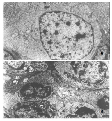
The apoptosis-inducing effect of SRCV was confirmed by electron microscopy. Compared with control group (Figure 1A), morphological changes indicative of apoptosis included cell shrinkage, nuclear chromatin condensation and peripheral shift of condensed chromatin to nuclear membrane or formation of crescent. In addition, the nuclear membrane was intact and there was little or no swelling of mitochondria or other organelles (Figure 1B).
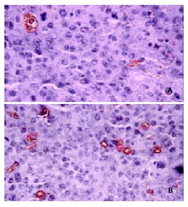
The apoptosis-inducing effect of SRCV was further confirmed by FCM and TUNEL. After treatment with SRCV, the apoptotic rate was significantly increased in the SRCV group compared with the control group (Table 2). In TUNEL assay, induction of apoptosis was represented by an increase in DNA fragments detected by a peroxidase reaction (Figure 2A), and the apoptotic cells in control tumors were scarcely scattered (Figure 2B).
Snake venoms have inhibitory effects on the growth of a variety of tumors in vitro and in vivo[20,21]. Markland et al[22] found that contortrostatin (CN), a homodimeric disintegrin from southern copperhead venom, inhibited dissemination ovarian cancer in a nude mouse model. According to Da Silva et al[23], Bothrops jararaca venom (BjV) had anti-tumor effects on Ehrlich ascites tumor (EAT) cells in vivo and in vitro.
Snake venom was also shown to induce apoptosis in tumors. Apoptosis, in contrast to necrosis, was an active process of gene-directed cellular suicide[24]. It has been clear that apoptosis is often upregulated in tumor by many anticancer drugs[25-32].
Since Araki et al[33] first described that hemorrhagic snake venoms induced apoptosis in vascular endothelial cells (VEC), data have been accumulated rapidly about apoptosis-inducing action of various snake venoms and their active components. In 1994, Strizhkov et al[34] reported that both neurotoxin II (NT II) from venom of Naja naja oxiana and 20-30 kDa proteins partially purified from pig brain (NTlm) cross-reacting with antibodies to NT II were cytotoxic to L929 and K562 tumor cells at concentrations of 10-6-10-8 M, and induced apoptosis in L929 and K562 cells in vitro. After that, L-amino acid oxidase (LAO) was found to induce apoptosis in human embryonic kidney cell line 293T[35] and human monocyte line MM6[36]. Recently, Araki et al[37] and Masuda et al[38] associated integrins with vascular apoptosis-inducing protein 1 (VAP1)-induced apoptosis. Data from Zhao et al[39] indicated that snake venom secreted phospholipase A2 (sPLA2) induced apoptosis in Mv1Lu cells in a dose- and time-dependent manner, and was associated with a rapid increase in intracellular ceramide level.
The clinical trial using snake venom and their active components have succeeded in cancer therapies, but its application was confined to the auxiliary treatment of patients in the late stage[40,41]. Its toxic and side effects were unavoidable. Great efforts have been made[42-45], but the problem remains unresolved. So a long-standing goal in snake venom therapy of cancer is to find a stable, low toxic, highly effective chemotherapeutic agent that selectively targets tumor cells. Based on this idea, we prepared the SRCV agent[8].
In our previous studies, the anti-tumor activity and apoptosis-inducing effect of SRCV were shown in vitro using HepG-2, HL-60 and human lung adenocarcinoma cell line, and no cytotoxicity was observed on human fetal lung fibroblast cells[46-48]. The results presented herein demonstrate that SRCV has inhibitory and apoptosis-inducing effects on implanted HepA tumors. Further studies are needed to identify the active components of SRCV and to elucidate their underlying mechanism.
Edited by Su Q and Wang XL
| 1. | Souza DH, Eugenio LM, Fletcher JE, Jiang MS, Garratt RC, Oliva G, Selistre-de-Araujo HS. Isolation and structural characterization of a cytotoxic L-amino acid oxidase from Agkistrodon contortrix laticinctus snake venom: preliminary crystallographic data. Arch Biochem Biophys. 1999;368:285-290. [RCA] [PubMed] [DOI] [Full Text] [Cited by in Crossref: 70] [Cited by in RCA: 66] [Article Influence: 2.5] [Reference Citation Analysis (0)] |
| 2. | Du XY, Clemetson KJ. Snake venom L-amino acid oxidases. Toxicon. 2002;40:659-665. [RCA] [PubMed] [DOI] [Full Text] [Cited by in Crossref: 266] [Cited by in RCA: 274] [Article Influence: 11.9] [Reference Citation Analysis (0)] |
| 3. | Corrêa MC, Maria DA, Moura-da-Silva AM, Pizzocaro KF, Ruiz IR. Inhibition of melanoma cells tumorigenicity by the snake venom toxin jararhagin. Toxicon. 2002;40:739-748. [RCA] [PubMed] [DOI] [Full Text] [Cited by in Crossref: 46] [Cited by in RCA: 43] [Article Influence: 1.9] [Reference Citation Analysis (0)] |
| 4. | Della Morte R, Squillacioti C, Garbi C, Derkinderen P, Belisario MA, Girault JA, Di Natale P, Nitsch L, Staiano N. Echistatin inhibits pp125FAK autophosphorylation, paxillin phosphorylation and pp125FAK-paxillin interaction in fibronectin-adherent melanoma cells. Eur J Biochem. 2000;267:5047-5054. [RCA] [PubMed] [DOI] [Full Text] [Cited by in Crossref: 19] [Cited by in RCA: 20] [Article Influence: 0.8] [Reference Citation Analysis (0)] |
| 5. | Zhou Q, Nakada MT, Brooks PC, Swenson SD, Ritter MR, Argounova S, Arnold C, Markland FS. Contortrostatin, a homodimeric disintegrin, binds to integrin alphavbeta5. Biochem Biophys Res Commun. 2000;267:350-355. [RCA] [PubMed] [DOI] [Full Text] [Cited by in Crossref: 32] [Cited by in RCA: 32] [Article Influence: 1.3] [Reference Citation Analysis (0)] |
| 6. | Kang IC, Lee YD, Kim DS. A novel disintegrin salmosin inhibits tumor angiogenesis. Cancer Res. 1999;59:3754-3760. [PubMed] |
| 7. | Abe Y, Shimoyama Y, Munakata H, Ito J, Nagata N, Ohtsuki K. Characterization of an apoptosis-inducing factor in Habu snake venom as a glycyrrhizin (GL)-binding protein potently inhibited by GL in vitro. Biol Pharm Bull. 1998;21:924-927. [RCA] [PubMed] [DOI] [Full Text] [Cited by in Crossref: 28] [Cited by in RCA: 25] [Article Influence: 0.9] [Reference Citation Analysis (0)] |
| 8. | Sun P, Xu C, Ren XD, Liu JJ, Li XH. Acute toxicity test of three Cobra Venom preparation and their inhibitory effect on transplanted hepatoma in mice. Jinan Daxue Xuebao. 2002;23:1-4. |
| 9. | Luo YR, Ye CL, Ren XD, Li HL, Zhong L. Inhibition of proliferation and apoptosis in HL60 cells induced by cobra venom serum. Zhongguo Yaolixue Tongbao. 2002;18:291-293. |
| 10. | Wang W, Qin SK, Chen BA, Chen HY. Experimental study on antitumor effect of arsenic trioxide in combination with cisplatin or doxorubicin on hepatocellular carcinoma. World J Gastroenterol. 2001;7:702-705. [PubMed] |
| 11. | Bi WX, Xu SD, Zhang PH, Kong F. Antitumoral activity of low density lipoprotein-aclacinomycin complex in mice bearing H (22) tumor. World J Gastroenterol. 2000;6:140-142. [PubMed] |
| 12. | Ryan KM, Ernst MK, Rice NR, Vousden KH. Role of NF-kappaB in p53-mediated programmed cell death. Nature. 2000;404:892-897. [RCA] [PubMed] [DOI] [Full Text] [Cited by in Crossref: 582] [Cited by in RCA: 599] [Article Influence: 24.0] [Reference Citation Analysis (0)] |
| 13. | Chun YJ, Park IC, Park MJ, Woo SH, Hong SI, Chung HY, Kim TH, Lee YS, Rhee CH, Lee SJ. Enhancement of radiation response in human cervical cancer cells in vitro and in vivo by arsenic trioxide (As2O3). FEBS Lett. 2002;519:195-200. [RCA] [PubMed] [DOI] [Full Text] [Cited by in Crossref: 52] [Cited by in RCA: 53] [Article Influence: 2.3] [Reference Citation Analysis (0)] |
| 14. | Lin SB, Wu LC, Huang SL, Hsu HL, Hsieh SH, Chi CW, Au LC. In vitro and in vivo suppression of growth of rat liver epithelial tumor cells by antisense oligonucleotide against protein kinase C-alpha. J Hepatol. 2000;33:601-608. [PubMed] [DOI] [Full Text] |
| 15. | Hou L, Li Y, Jia YH, Wang B, Xin Y, Ling MY, Lü S. Molecular mechanism about lymphogenous metastasis of hepatocarcinoma cells in mice. World J Gastroenterol. 2001;7:532-536. [PubMed] |
| 16. | Tian G, Yu JP, Luo HS, Yu BP, Yue H, Li JY, Mei Q. Effect of nimesulide on proliferation and apoptosis of human hepatoma SMMC-7721 cells. World J Gastroenterol. 2002;8:483-487. [PubMed] |
| 17. | Cui RT, Cai G, Yin ZB, Cheng Y, Yang QH, Tian T. Transretinoic acid inhibits rats gastric epithelial dysplasia induced by N-methyl-N-nitro-N-nitrosoguanidine: influences on cell apoptosis and expression of its regulatory genes. World J Gastroenterol. 2001;7:394-398. [PubMed] |
| 18. | Barnett KT, Fokum FD, Malafa MP. Vitamin E succinate inhibits colon cancer liver metastases. J Surg Res. 2002;106:292-298. [RCA] [PubMed] [DOI] [Full Text] [Cited by in Crossref: 65] [Cited by in RCA: 63] [Article Influence: 2.7] [Reference Citation Analysis (0)] |
| 19. | Zhang XL, Liu L, Jiang HQ. Salvia miltiorrhiza monomer IH764-3 induces hepatic stellate cell apoptosis via caspase-3 activation. World J Gastroenterol. 2002;8:515-519. [PubMed] |
| 20. | Zhou Q, Sherwin RP, Parrish C, Richters V, Groshen SG, Tsao-Wei D, Markland FS. Contortrostatin, a dimeric disintegrin from Agkistrodon contortrix contortrix, inhibits breast cancer progression. Breast Cancer Res Treat. 2000;61:249-260. [RCA] [PubMed] [DOI] [Full Text] [Cited by in Crossref: 66] [Cited by in RCA: 73] [Article Influence: 2.9] [Reference Citation Analysis (0)] |
| 21. | Yeh CH, Peng HC, Yang RS, Huang TF. Rhodostomin, a snake venom disintegrin, inhibits angiogenesis elicited by basic fibroblast growth factor and suppresses tumor growth by a selective alpha (v)beta (3) blockade of endothelial cells. Mol Pharmacol. 2001;59:1333-1342. [PubMed] |
| 22. | Markland FS, Shieh K, Zhou Q, Golubkov V, Sherwin RP, Richters V, Sposto R. A novel snake venom disintegrin that inhibits human ovarian cancer dissemination and angiogenesis in an orthotopic nude mouse model. Haemostasis. 2001;31:183-191. [RCA] [PubMed] [DOI] [Full Text] [Cited by in Crossref: 5] [Cited by in RCA: 20] [Article Influence: 0.8] [Reference Citation Analysis (0)] |
| 23. | da Silva RJ, da Silva MG, Vilela LC, Fecchio D. Antitumor effect of Bothrops jararaca venom. Mediators Inflamm. 2002;11:99-104. [RCA] [PubMed] [DOI] [Full Text] [Full Text (PDF)] [Cited by in Crossref: 18] [Cited by in RCA: 20] [Article Influence: 0.9] [Reference Citation Analysis (0)] |
| 24. | Yu CL, Tsai MH. Fetal fetuin selectively induces apoptosis in cancer cell lines and shows anti-cancer activity in tumor animal models. Cancer Lett. 2001;166:173-184. [RCA] [PubMed] [DOI] [Full Text] [Cited by in Crossref: 11] [Cited by in RCA: 12] [Article Influence: 0.5] [Reference Citation Analysis (0)] |
| 25. | Wu K, Zhao Y, Liu BH, Li Y, Liu F, Guo J, Yu WP. RRR-alpha-tocopheryl succinate inhibits human gastric cancer SGC-7901 cell growth by inducing apoptosis and DNA synthesis arrest. World J Gastroenterol. 2002;8:26-30. [PubMed] |
| 26. | Panichakul T, Wanun T, Reutrakul V, Sirisinha S. Synergistic cytotoxicity and apoptosis induced in human cholangiocarcinoma cell lines by a combined treatment with tumor necrosis factor-alpha (TNF-alpha) and triptolide. Asian Pac J Allergy immunol. 2002;20:167-173. [PubMed] |
| 27. | Zhang XL, Liu L, Jiang HQ. Salvia miltiorrhiza monomer IH764-3 induces hepatic stellate cell apoptosis via caspase-3 activation. World J Gastroenterol. 2002;8:515-519. [PubMed] |
| 28. | Sun ZX, Ma QW, Zhao TD, Wei YL, Wang GS, Li JS. Apoptosis induced by norcantharidin in human tumor cells. World J Gastroenterol. 2000;6:263-265. [PubMed] |
| 29. | Zhang C, Gong Y, Ma H, An C, Chen D, Chen ZL. Reactive oxygen species involved in trichosanthin-induced apoptosis of human choriocarcinoma cells. Biochem J. 2001;355:653-661. [RCA] [PubMed] [DOI] [Full Text] [Cited by in Crossref: 66] [Cited by in RCA: 71] [Article Influence: 3.0] [Reference Citation Analysis (0)] |
| 30. | Wang ZM, Hu J, Zhou D, Xu ZY, Panasci LC, Chen ZP. Trichostatin A inhibits proliferation and induces expression of p21WAF and p27 in human brain tumor cell lines. Ai Zheng. 2002;21:1100-1105. [PubMed] |
| 31. | Zhao AG, Zhao HL, Jin XJ, Yang JK, Tang LD. Effects of Chinese Jianpi herbs on cell apoptosis and related gene expression in human gastric cancer grafted onto nude mice. World J Gastroenterol. 2002;8:792-796. [PubMed] |
| 32. | Tu SP, Zhong J, Tan JH, Jiang XH, Qiao MM, Wu YX, Jiang SH. Induction of apoptosis by arsenic trioxide and hydroxy camptothecin in gastriccancer cells in vitro. World J Gastroenterol. 2000;6:532-539. [PubMed] |
| 33. | Du XY, Clemetson KJ. Snake venom L-amino acid oxidases. Toxicon. 2002;40:659-665. [RCA] [PubMed] [DOI] [Full Text] [Cited by in Crossref: 266] [Cited by in RCA: 274] [Article Influence: 11.9] [Reference Citation Analysis (0)] |
| 34. | Strizhkov BN, Blishchenko EYu DK, Karelin AA. Both neurotoxin II from venom of Naja naja oxiana and its endogenous analogue induce apoptosis in tumor cells. FEBS Lett. 1994;340:22-24. [RCA] [PubMed] [DOI] [Full Text] [Cited by in Crossref: 15] [Cited by in RCA: 17] [Article Influence: 0.5] [Reference Citation Analysis (0)] |
| 35. | Torii S, Yamane K, Mashima T, Haga N, Yamamoto K, Fox JW, Naito M, Tsuruo T. Molecular cloning and functional analysis of apoxin I, a snake venom-derived apoptosis-inducing factor with L-amino acid oxidase activity. Biochemistry. 2000;39:3197-3205. [RCA] [PubMed] [DOI] [Full Text] [Cited by in Crossref: 81] [Cited by in RCA: 79] [Article Influence: 3.2] [Reference Citation Analysis (0)] |
| 36. | Ali SA, Stoeva S, Abbasi A, Alam JM, Kayed R, Faigle M, Neumeister B, Voelter W. Isolation, structural, and functional characterization of an apoptosis-inducing L-amino acid oxidase from leaf-nosed viper (Eristocophis macmahoni) snake venom. Arch Biochem Biophys. 2000;384:216-226. [RCA] [PubMed] [DOI] [Full Text] [Cited by in Crossref: 76] [Cited by in RCA: 78] [Article Influence: 3.3] [Reference Citation Analysis (0)] |
| 37. | Araki S, Masuda S, Maeda H, Ying MJ, Hayashi H. Involvement of specific integrins in apoptosis induced by vascular apoptosis-inducing protein 1. Toxicon. 2002;40:535-542. [RCA] [PubMed] [DOI] [Full Text] [Cited by in Crossref: 39] [Cited by in RCA: 32] [Article Influence: 1.4] [Reference Citation Analysis (0)] |
| 38. | Masuda S, Ohta T, Kaji K, Fox JW, Hayashi H, Araki S. cDNA cloning and characterization of vascular apoptosis-inducing protein 1. Biochem Biophys Res Commun. 2000;278:197-204. [RCA] [PubMed] [DOI] [Full Text] [Cited by in Crossref: 49] [Cited by in RCA: 46] [Article Influence: 1.8] [Reference Citation Analysis (0)] |
| 39. | Zhao S, Du XY, Chai MQ, Chen JS, Zhou YC, Song JG. Secretory phospholipase A (2) induces apoptosis via a mechanism involving ceramide generation. Biochim Biophys Acta. 2002;1581:75-88. [RCA] [PubMed] [DOI] [Full Text] [Cited by in Crossref: 21] [Cited by in RCA: 19] [Article Influence: 0.8] [Reference Citation Analysis (0)] |
| 40. | Cura JE, Blanzaco DP, Brisson C, Cura MA, Cabrol R, Larrateguy L, Mendez C, Sechi JC, Silveira JS, Theiller E. Phase I and pharmacokinetics study of crotoxin (cytotoxic PLA (2), NSC-624244) in patients with advanced cancer. Clin Cancer Res. 2002;8:1033-1041. [PubMed] |
| 41. | Costa LA, Miles HA, Diez RA, Araujo CE, Coni Molina CM, Cervellino JC. Phase I study of VRCTC-310, a purified phospholipase A2 purified from snake venom, in patients with refractory cancer: safety and pharmacokinetic data. Anticancer Drugs. 1997;8:829-834. [RCA] [PubMed] [DOI] [Full Text] [Cited by in Crossref: 21] [Cited by in RCA: 22] [Article Influence: 0.8] [Reference Citation Analysis (0)] |
| 42. | Fu Q, Gowda DC. Carbohydrate-directed conjugation of cobra venom factor to antibody by selective derivatization of the terminal galactose residues. Bioconjug Chem. 2001;12:271-279. [RCA] [PubMed] [DOI] [Full Text] [Cited by in Crossref: 7] [Cited by in RCA: 7] [Article Influence: 0.3] [Reference Citation Analysis (0)] |
| 43. | Wang XM, Huang SJ. The selective cytotoxicity of cobra venom factor immunoconjugate on cultured human nasopharyngeal carcinoma cell line. Hum Exp Toxicol. 1999;18:71-76. [RCA] [PubMed] [DOI] [Full Text] [Cited by in Crossref: 3] [Cited by in RCA: 3] [Article Influence: 0.1] [Reference Citation Analysis (0)] |
| 44. | Schmitmeier S, Markland FS, Chen TC. Anti-invasive effect of contortrostatin, a snake venom disintegrin, and TNF-alpha on malignant glioma cells. Anticancer Res. 2000;20:4227-4233. [PubMed] |
| 45. | Juhl H, Petrella EC, Cheung NK, Bredehorst R, Vogel CW. Additive cytotoxicity of different monoclonal antibody-cobra venom factor conjugates for human neuroblastoma cells. Immunobiology. 1997;197:444-459. [RCA] [PubMed] [DOI] [Full Text] [Cited by in Crossref: 25] [Cited by in RCA: 19] [Article Influence: 0.7] [Reference Citation Analysis (0)] |
| 46. | Li HL, Ren XD, Luo YR, Ye CL, Zhang HW. Effect of serum preparation derived from rabbits after the gastrolavage with Naja naja atra venom on human lung adenocarcinoma cell line. Jinan Daxue Xuebao. 2001;22:1-5. |
| 47. | Li HL, Ren XD, Luo YR, Ye CL, Zhang HW. Effect of the oral administration with serum preparation of Naja naja atra venom on apoptosis in HepG2 cells. Zhongguo Binglishengli Zazhi. 2002;18:199-200. |
| 48. | Li HL, Sun P, Ren XD, Luo YR, Ye CL, Zhang HW. A antihepatoc-arcinoma activity of the oral serum preparation of Naja naja atra venom. Health Science J Macao. 2002;2:59-61. |


