Published online Apr 15, 2001. doi: 10.3748/wjg.v7.i2.235
Revised: February 27, 2001
Accepted: March 1, 2001
Published online: April 15, 2001
AIM: Of this investigation is to reveal the damage to peripheral blood lymphocytes (PBL) DNA in the patients with chronic liver diseases.
MATERIALS AND METHODS: Sixteen-nine patients with chronic liver diseases (37 patients with chronic viral hepatitis, 2 patients with liver cirrhosis of mixed etiology (alcohol + virus G), 30 women with primary biliary cirrhosis-PBC) were examined. The condition of DNA structure of PBL was measured by the fluorescence analysis of DNA unwinding (FADU) technique with modification. Changes of fluorescence (in %) reflected the DNA distractions degree (the presence of DNA single-stranded breaks and alkalinelabile sights).
RESULTS AND CONCLUSION: The quantity of DNA single-stranded breaks and alkalinelabile sights in DNA in all patients with chronic viral hepatitis didn’t differ from the control group, excluding the patients with chronic hepatitis (CH) C + G. Patients with HGV and TTV monoinfection had demonstrated the increase of the DNA single-stranded breaks PBL quantity. This fact may be connected with hypothesis about the viruses replication in white blood cells discussed in the literature. Tendency to increase quantity of DNA PBL damages in the patients with primary biliary cirrhosis (PBC) accordingly to the alkaline phosphatase activity increase was revealed. Significant decrease of the DNA single-stranded breaks and alkalinelabile sights in the PBC patients that were treated with prednison was demonstrated. Probably, the tendency to increase the quantity of DNA single stranded breaks and alkalinelabile sights in lymphocytes of the PBC patients was depended on the surplus of the blood bile acid content.
- Citation: Reshetnyak VI, Sharafanova TI, Ilchenko LU, Golovanova EV, Poroshenko GG. Peripheral blood lymphocytes DNA in patients with chronic liver diseases. World J Gastroenterol 2001; 7(2): 235-237
- URL: https://www.wjgnet.com/1007-9327/full/v7/i2/235.htm
- DOI: https://dx.doi.org/10.3748/wjg.v7.i2.235
The last years investigations demonstrated that chronic viral hepatitis are the systemic infections[1,2]. The replication of viruses hepatitis B (HBV), C (HCV) was revealed in mononuclear blood cells, bone marrow, lien and other organs. This fact accounts for polymorphism clinical signs of these infections and sometimes-unsuccessful treatment with interferon’s therapy.
The role of recently revealed hepatitis G virus (HGV) for the autoimmune processes development is being discussed[3]. Phenomenon of viral immune supervision “avoid” is associated with disturbances of infected lymphocytes and monocytes immune control functions. Viral replication in blood cells with nucleuses may lead to the damage of lymphocytes genetic apparatus and the beginning of immunopathological reactions.
The immunocompetent cells condition plays significant role in the development of autoimmune process in the patients with PBC.THE AIM of this investigation is to reveal the damage to peripheral blood lymphocytes (PBL) DNA in the patients with chronic liver diseases.
We studied 69 patients with chronic liver diseases: 43 females and 26 male, mean age ± SD: 40.8 ± 17.7 years (range 16-77 years). The study group consisted of 37 patients with chronic viral hepatitis, 2 patients with liver cirrhosis of mixed etiology (alcohol + virus G), 30 woman with PBC. Control group was formed of 10 healthy volunteers (5 females and 5 males).
The liver functional state was estimated with cytolysis enzymes activity (alanine and asparagine aminotransferases), indexes of cholestasis syndrome (activity of alkaline phosphatase, gammaglutamiltranspeptidase, and contents of cholesterol, bilirubin).
Hepatitis B markers (HBsAg, HBsAb, HBeAg, HBeAb, HBcAbsum, HBcAb IgM, HBV DNA), hepatitis C markers (HCVAb IgG, HCVAb IgM, HCV RNA) hepatitis G markers (HGV RNA) and hepatitis TTV markers (TTV DNA) were revealed with immunoferment analysis method (IFA) and with polymerase chain reaction(PCR). The immunoglobuline’s levels (IgM, IgG, IgA) were determined with IFA.
The liver biopsy was done to all patients with PBC and the main part of the patients with chronic hepatitis.
Lymphocytes were obtained from 20 mL of peripheral blood (obtained by venepuncture from healthy donors and patients with chronic liver disease) by centrifuge with the presence of Fikoll-pak solution during 30 min with 400 × g.
The condition of DNA structure of PBL was measured by the fluorescence analysis of DNA unwinding (FADU) technique as indicated by Birnboim H.C. and Jevcak J.J. (1981) with modification[4,5]. Method was created for alkalinelable sights and DNA single-stranded breaks registration. Suspension with 2 × 10⁶ cells/mL was lysised. Lysate was treated with ethidium bromide, which selectively binds to double-stranded (ds) DNA. Ethidium bromide with DNA formed fluorescence complexes.
Each lysate was divided equally into 3 sets of tubes: ① a sample (P) used for evaluating total fluorescence of native DNA; ② a blank sample (B) in which DNA was completely unwound by alkaline and mechanical destruction; ③ a sample (T) used to estimate alkalinelabile sights and DNA single-stranded breaks which was obtained by alkaline. The percentage of dsDNA (D) was determined from the fluorescence of tubes P, B and T by equation: D = (P - B)/(T - B) × 100%.
The fluorescence was read with excitation wavelength 520 nm and an emission wavelength of 590 nm, and slit width 8 nm by a Jasco FP-550 spectrofluorimeter.
Changes of fluorescence (in %) reflected the DNA distractions degree (the presence of DNA single-stranded breaks and alkalinelabile sights).
Conventional methods were used for calculation of means and standard deviations (SD). Results are shown as means ± SD. For skewed variables, non-parametric tests were used for comparisons between the groups (Mann-Whitney U-test), whereas Student’s t-test was used for normally distributed variables. In all cases, P values less than 0.05 were considered significant.
There were 37 patients with chronic viral hepatitis in our study. Chronic hepatitis B was revealed in 5 patients, chronic hepatitis C in 11 patients, chronic hepatitis B + C-in 1 patients, chronic hepatitis G-in 8 patients, chronic hepatitis TTV-in 4 patients, chronic hepatitis B + G-in 2 patients, chronic hepatitis C + G-in 3 patients, and chronic hepatitis B + C + G-in 3 patients.
Cirrhosis of mixed etiology (alcohol+virus G) was revealed in 2 of 69 studied patients. Replication of viruses was revealed in all patients with chronic viral hepatitis and liver cirrhosis with mixed etiology.
PBC was determined in 30 of 69 studied patients. The 1st-2 nd stage of PBC determined in 10 patients, the 3 rd stage of PBC was revealed in 14 patients and the 4th stage of PBC was revealed in 6 patients (accordingly to morphological H.Popper’s classification, 1970)[6].
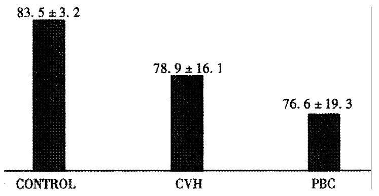
The percentage of ds DNA, D, in control group was 83.5 ± 3.2. PBL DNA damages in patients with CH and PBC did not significantly differ from control group (Figure 1).
The tendency to the increase of alkalinelabile sights and DNA single-stranded breaks in the patients with chronic liver diseases was determined.
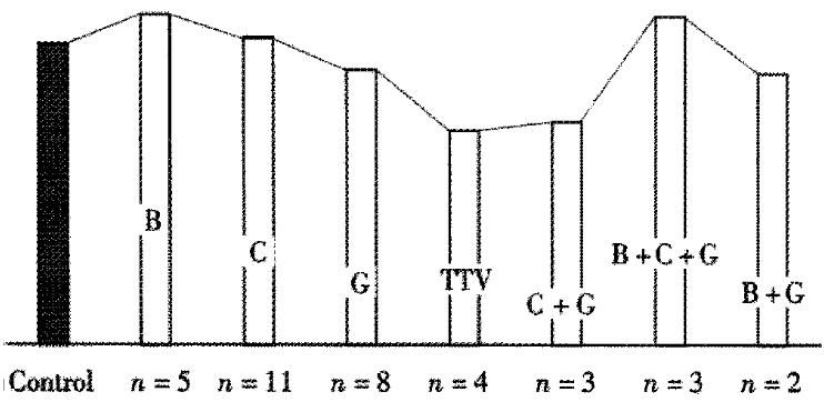
Figure 2 shows diagram of DNA PBL content in the patients with viral CH. The quantity of DNA single-stranded breaks and alkalinelabile sights in all patients of this group didn’t differ from the control group, excluding the patients with CH C+G (-x±s: D = 65.3% ± 12.9%, P < 0.05).
Apparently the viruses C and B didn’t possess a significant destructive effect on DNA PBL of the patients with chronic disease. But the patients with HGV and TTV monoinfection had demonstrated the tendency of increase of the DNA single-stranded breaks PBL quantity. This fact may be connected with hypothesis about the viruses replication in white blood cells discussed in the literature[3].
Increase of the HGV destructive effect on the DNA was revealed in the patients with chronic hepatitis of mixed (HCV + HGV) etiology (quantity of alkalinelabile sights and DNA single-stranded breaks is increased) (Figure 2).
Percentage of ds DNA in group of the patients with mixed HBV + HCV + HGV infections was decreased (D = 63.5 ± 20.7). Correlation between DNA structure change and hyperfermentemia degree was not revealed in the patients with different activity of chronic hepatitis.
So, monoinfection with HBV and HCV didn’t significantly influence on PBL DNA structure in the patients with chronic disease. Monoinfection with HGV and TTV leads to increase of DNA single-stranded breaks in human PBL. Possibly, HCV increases virus G destructive effect on lymphocytes DNA structure.
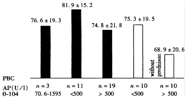
Content of ds DNA in PBL in depending on cholestasis degree (activity of alkaline phosphatase, content of cholesterol and bilirubin), degree of histological process and used treatment was examined in the patients with PBC. Tendency to increase quantity of DNA PBL damages in the patients with PBC according to the alkaline phosphatase activity increase (Daph < 500un/ = 81.9% ± 15.2%, n = 11; D aph > 500un/L = 74.8% ± 21.8%, n = 19) (Figure 3) was revealed.
The same data was demonstrated in 20 PBC patients without prednison treatment: percentage of ds DNA was 75.5% ± 19.5% in 10 PBC patients with alkaline phosphatase activity < 500 u/L, and D = 68. 9% ± 20.6% in 10 PBC patients with alkaline phosphatase activity > 500 u/L (Figure 3) . Dependence on ds DNA content according to other indexes of cholestasis and degree of histological changes was not demonstrated.
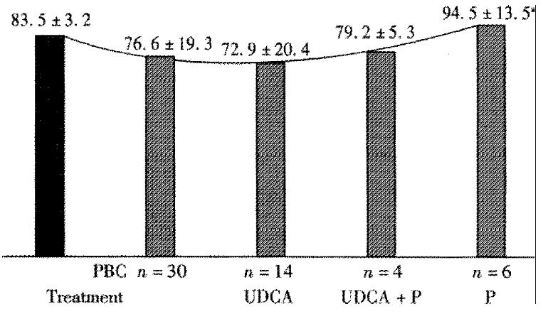
All PBC patients were divided into 4 groups according to the treatment methods: 6 patients were treated with prednison, 14 patients were treated with ursodeoxycholic acid (UDCA), 4 patients were treated with prednison and UDCA simultaneously, and 6 patients were treated with metabolic drug. The UDCA was not increased of D (Figure 4). Significant decrease of the DNA single-stranded breaks and alkalinelabile sights in the PBC patients, that were treated with prednison (-x±s: D = 94.5% ± 13.5%, n = 6, P < 0.05, versus control D = 72.2% ± 19.9%, n = 20, P < 0.05) was revealed (Figure 4).
The percentage of ds DNA, D, in lymphocytes in the PBC patients that were treated with prednison and UDCA simultaneously was 79.2% ± 5.3% (Figure 4).
Index D in the patients that were treated with UDCA, prednison and prednison with UDCA simultaneously had testified the suppression by bile acids the DNA reparation that was depended on prednison.
Probably, the tendency to increase the quantity of DNA single-stranded breaks and alkalinelabile sights in lymphocytes of the PBC patients was depended on the surplus of the blood bile acid content.
Grant source is the budget of the Central Research Institute of Gastroenterology that was supported by Department of Moscow Public Health.
Edited by Ma JY
| 1. | Aprosina ZG, Serov VV. [Chronic viral diseases of the liver: their patho- and morphogenesis and clinical characteristics]. Ter Arkh. 1995;67:77-80. [PubMed] |
| 2. | Aprosina ZG, Borisova VV, Krel' PE, Serov VV, Sklianskaia OA. [The unique course of a chronic generalized infection with the hepatitis B virus (a clinico-morphological observation)]. Ter Arkh. 1996;68:16-19. [PubMed] |
| 3. | Tribl B, Schöniger-Hekele M, Petermann D, Bakos S, Penner E, Müller C. Prevalence of GBV-C/HGV-RNA, virus genotypes, and anti-E2 antibodies in autoimmune hepatitis. Am J Gastroenterol. 1999;94:3336-3340. [RCA] [PubMed] [DOI] [Full Text] [Cited by in Crossref: 6] [Cited by in RCA: 7] [Article Influence: 0.3] [Reference Citation Analysis (0)] |
| 4. | Birnboim HC, Jevcak JJ. Fluorometric method for rapid detection of DNA strand breaks in human white blood cells produced by low doses of radiation. Cancer Res. 1981;41:1889-1892. [PubMed] |
| 5. | Thierry D, Rigaud O, Duranton I, Moustacchi E, Magdelenat H. Quantitative measurement of DNA strand breaks and repair in gamma-irradiated human leukocytes from normal and ataxia telangiectasia donors. Radiat Res. 1985;102:347-358. [RCA] [PubMed] [DOI] [Full Text] [Cited by in Crossref: 24] [Cited by in RCA: 24] [Article Influence: 0.6] [Reference Citation Analysis (0)] |
| 6. | Popper H, Schaffner F. Nonsuppurative destructive chronic cholangitis and chronic hepatitis. Prog Liver Dis. 1970;3:336-354. [PubMed] |


