Copyright
©The Author(s) 1999.
World J Gastroenterol. Dec 15, 1999; 5(6): 492-505
Published online Dec 15, 1999. doi: 10.3748/wjg.v5.i6.492
Published online Dec 15, 1999. doi: 10.3748/wjg.v5.i6.492
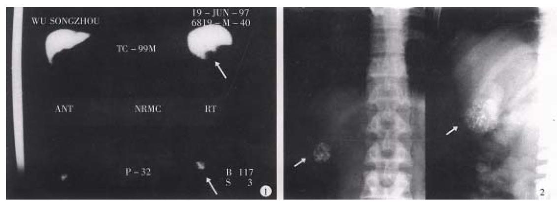
Figure 10 Male, aged 41, hospital no.
207089, clinical diagnosis: uremia, right hepatic small carcinoma (4 cm × 3 cm), with A FP > 400 μg/L treated by regimen A for 2 courses with interval of 3 months, resulted in AFP restored to normal level and decreased tumor size over 50%. 1, Colloidal SPECT imaging showed the space occupying lesion in the right liver lobe (as shown by arrow). 2, Comparing the tumor size before and after treatment of plain film (as arrow shown), the patient died from renal failure 26 months after operation.
- Citation: Liu L, Jiang Z, Teng GJ, Song JZ, Zhang DS, Guo QM, Fang W, He SC, Guo JH. Clinical and experimental study on regional administration of phosphorus 32 glass microspheres in treating hepatic carcinoma. World J Gastroenterol 1999; 5(6): 492-505
- URL: https://www.wjgnet.com/1007-9327/full/v5/i6/492.htm
- DOI: https://dx.doi.org/10.3748/wjg.v5.i6.492


