Published online Sep 15, 1997. doi: 10.3748/wjg.v3.i3.163
Revised: October 13, 1996
Accepted: November 19, 1996
Published online: September 15, 1997
AIM: To study P21 and carcinoembryonic antigen (CEA) expression and to measure argyrophilic nucleolar organizer region (AgNOR) counts in various lesions of colonic mucosa and the mechanism of carcinogenesis.
METHODS: Thirty-eight male Wistar rats were injected with dimethylhydrazine (DMH) once a week for 25 wk. P21 and CEA expression was detected by immunohistochemical methods, and AgNOR was counted by silver staining paraffin sections from various colonic lesions.
RESULTS: The incidence of colonic carcinoma in DMH-treated rats was 71.05% (27/38), and lymph node metastasis occurred in six rats. Immunohistochemical studies showed that P21 was primarily expressed in dysplasia and carcinomas, while CEA was expressed in carcinomas and metastatic tumors. AgNOR counts were higher in dysplasia and carcinomas. There were significant differences in P21 and CEA expression between benign and malignant lesions (P < 0.05). The difference in AgNOR counts was also significant between normal and dysplastic tissues, and between dysplasia and malignant lesions (P < 0.05).
CONCLUSION: Dysplasia is a premalignant change of colonic carcinoma. The detection of P21 via immunohistochemistry and AgNOR counting may be an important clinical screening technique for colon carcinoma and premalignant lesions.
- Citation: Zhang ZG, Wu JY, Fu XD, Gu DK, Fang F. P21 and CEA expression and AgNOR counts in dimethylhydrazine-induced colon carcinoma in rats. World J Gastroenterol 1997; 3(3): 163-165
- URL: https://www.wjgnet.com/1007-9327/full/v3/i3/163.htm
- DOI: https://dx.doi.org/10.3748/wjg.v3.i3.163
The transformation-inducing genes most frequently detected in solid human tumors are members of the Ras family of oncogenes. It has been suggested that P21 overexpression in the early neoplasm stage may play an important role in carcinogenesis[1,2]. In this study, we examined P21 and CEA expression and quantified nuclear silver stain of the granules of nucleolus organizer region (AgNOR) in 1.2 dimethylhydrazine (DMH)-induced colonic carcinoma in rats to study the carcinogenesis mechanism.
Fifty male Wistar rats weighing approximately 170 g were randomly divided into a test group (T group, n = 40) and control group (C group, n = 10). Rats in the T group were subcutaneously injected with 20 mg/kg body weight DMH once a week. Rats in the C group rats were given 0.9% NaCl solution at the same time. DMH administration lasted 20 wk. At the 10th, 15th, and 18th week, one rat was sacrificed in each group, and 10 were sacrificed at the 20th week. The rest were continually fed with standard diet until the 25th week, when they were all sacrificed. Intestinal tissues were fixed in 10% neutral formalin.
Tissue specimens were collected from the tumor, adjacent tissues and normal colonic mucosa in T group rats, and normal colon mucosa were collected from the C group. Serial paraffin sections were cut at 4 μm thickness and were stained with hematoxylin and eosin (HE). The ABC immunohistochemical method was used for P21 staining, and the PAP method was used for CAE staining. The working dilution of the anti-P21 monoclonal antibody (Huamei Company) was 1:40, and 1:100 for the anti-CEA antibody (DAKO Company). PBS was used as a negative control.
A one-step silver staining method was used for AgNOR detection. The observation method and statistical standard were performed as described previously[3].
Of the 38 rats in the T group (two rats unexpectedly died during the experiment and were excluded), 27 developed colonic carcinoma with a total of 34 masses. The incidence of carcinogenesis was 71.05% (27/38). Various colonic mucosal lesions are summarized in Table 1. The tumors were 1-13 mm in diameter and more frequently occurred in the distal colon. Most tumors appeared nodular in shape. Six rats developed mesenteric lymph node metastasis, and two developed gastric and pulmonary metastases.
| Lesions | n | Incidence (%) | Number of mass |
| Well-differentiated adenocarcinoma | 17 | 44.74 | 20 |
| Undifferentiated adenocarcinoma | 3 | 7.89 | 5 |
| Mucinous adenocarcinoma | 7 | 18.42 | 9 |
| No tumor | 11 | ||
| Total | 38 | 71.05 | 34 |
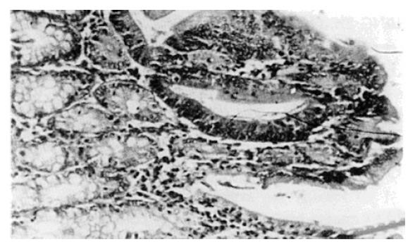
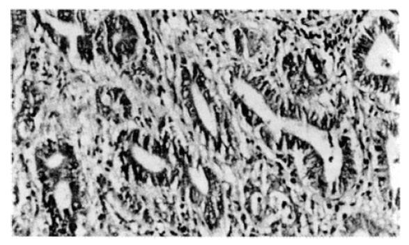
T group rats exhibited inflammation and atypical dysplasia of the colonic mucosa at the 10th, 15th, and 18th week (Figure 1 and Figure 2). We observed adenomas and adenocarcinomas after 20 wk with metastasis (Table 2). We never observed dysplasia or tumor development in the control group.
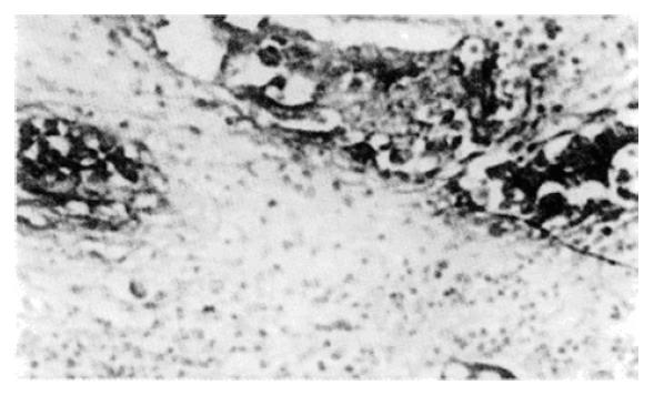
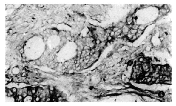
P21 and CEA staining of colonic lesions is summarized in Table 3. Normal and inflammatory mucosa were negative for expression. P21 was weakly expressed in adenomas and primarily expressed in dysplasia and carcinoma (Figure 3), while CEA was expressed in dysplasia, adenocarcinoma and metastasis (Figure 4). The difference in P21 and CEA expression between benign and malignant colonic mucosal lesions in DMH-treated rats was statistically significant (P < 0.05).
| Histological types | n | p21 (%) | CEA (%) |
| Benign | |||
| Normal epithelial cell | 10 | 0 (0.0) | 0 (0.0) |
| Adenoma | 3 | 1 (33.3) | 0 (0.0) |
| Dysplasia | 10 | 6 (60.0) | 3 (30.0) |
| Malignant | |||
| Well-differentiated adenocarcinoma | 20 | 14 (70.0) | 16 (80.0) |
| Undifferentiated adenocarcinoma | 5 | 1 (20.0) | 4 (80.0) |
| Mucinous adenocarcinoma | 9 | 7 (77.8) | 6 (66.7) |
The AgNOR counts in various colonic mucosa lesions were as follows: adenoma 2.7 ± 0.18, dysplasia 3.2 ± 0.21, and adenocarcinoma 4.6 ± 0.26, and normal epithelial cell 1.45 ± 0.08. In colonic adenocarcinoma, the AgNOR granules not only increased in number, but also became larger and irregular. The AgNOR counts were statistically significantly different between normal mucosa and dysplasia, as well as dysplasia and malignant tissue (P < 0.05). However, among the various colonic carcinomas, the differences were not statistically significant (P > 0.05).
We treated rats with DMH to induce colonic carcinoma and observed the sequential changes in the colonic mucosa. The marked dysplasia in the epithelial cells initially involved one or two glands and was localized to the upper 1/3 of the mucosal layer (Figure 1). As the experiment progressed, the number of atypical glands increased, and mucosal noduli formed. Cell heterogeneity also increased, and ultimately led to colonic adenocarcinoma with invasion and metastasis, strongly suggesting that dysplasia is a premalignant lesion for colonic carcinoma.
The activation of oncogenes and P21 overexpression might be critical for malignant cell transformation[4]. P21 can inhibit the intercellular signal transmission, leading to loss of controlled cell mitosis. Therefore, P21 overexpression is an important sign of carcinogenesis[5]. In our study, we found that P21 protein was expressed in both dysplasia and adenocarcinoma. These data suggest that Ras may exert its effects on cell growth as early as the dysplasia stage to further promote cell transformation. The results support the hypothesis that dysplasia is a premalignant lesion of colonic carcinoma; therefore, ras P21 expression could be an early diagnostic indicator for premalignant lesions. CEA expression is another major marker in colonic carcinoma diagnosis. In DMH-induced colon carcinoma, dysplasia presented with P21 overexpression and increased AgNOR counts, while CEA was only expressed in carcinoma and metastasis. In metastasis, the AgNOR granules increased and clustered, which is a typical indicator of the malignant colonic carcinoma phenotype. We suggest that P21 and CEA are two proteins expressed in colon carcinoma at different stages of differentiation. In conclusion, P21 immunohistochemical detection and AgNOR quantification important for clinical screening of colon carcinoma and premalignant lesions.
Zhi-Gang Zhang, works in the Department of Pathology, Shanghai Medical University, and has published ten papers and one book.
*This project was funded by the Youth Research Fund of Shanghai Municipal Health Bureau (131914Y5).
Original title:
S- Editor: Filipodia L- Editor: Jennifer E- Editor: Hu S
| 1. | Carneiro F, David L, Sunkel C, Lopes C, Sobrinho-Simões M. Immunohistochemical analysis of ras oncogene p21 product in human gastric carcinomas and their adjacent mucosas. Pathol Res Pract. 1992;188:263-272. [RCA] [PubMed] [DOI] [Full Text] [Cited by in Crossref: 7] [Cited by in RCA: 7] [Article Influence: 0.2] [Reference Citation Analysis (0)] |
| 2. | Viola MV, Fromowitz F, Oravez S, Deb S, Schlom J. ras Oncogene p21 expression is increased in premalignant lesions and high grade bladder carcinoma. J Exp Med. 1985;161:1213-1218. [RCA] [PubMed] [DOI] [Full Text] [Full Text (PDF)] [Cited by in Crossref: 125] [Cited by in RCA: 122] [Article Influence: 3.1] [Reference Citation Analysis (0)] |
| 3. | Zhang ZG, Zhai WR, Zhang JM, Zhong TL. Study on CEA with immunohistochemistry and AgNOR count in breast carcinoma (in Chinese with English abstract). Chinese Journal of Histochemistry Cytochemistry. 1994;3:374-378. |
| 4. | He KL. Oncogene and growth factor (in Chinese). In: Tang ZY, eds Modern oncology, Shanghai: Shanghai Medical University Publisher 1993; 71-75. |
| 5. | Spandidos DA, Kerr IB. Elevated expression of the human ras oncogene family in premalignant and malignant tumours of the colorectum. Br J Cancer. 1984;49:681-688. [RCA] [PubMed] [DOI] [Full Text] [Full Text (PDF)] [Cited by in Crossref: 135] [Cited by in RCA: 137] [Article Influence: 3.3] [Reference Citation Analysis (0)] |


