Copyright
©The Author(s) 2020.
World J Gastroenterol. Nov 14, 2020; 26(42): 6698-6705
Published online Nov 14, 2020. doi: 10.3748/wjg.v26.i42.6698
Published online Nov 14, 2020. doi: 10.3748/wjg.v26.i42.6698
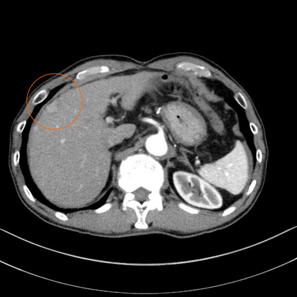
Figure 1 Abdominal contrast-enhanced computed tomography before the first surgery.
Arterial phase of abdominal contrast-enhanced computed tomography before the first surgery showed a tumor nodule 20 mm in diameter with early staining located in segment 8 of the liver (orange circle).
- Citation: Mashiko T, Masuoka Y, Nakano A, Tsuruya K, Hirose S, Hirabayashi K, Kagawa T, Nakagohri T. Intussusception due to hematogenous metastasis of hepatocellular carcinoma to the small intestine: A case report. World J Gastroenterol 2020; 26(42): 6698-6705
- URL: https://www.wjgnet.com/1007-9327/full/v26/i42/6698.htm
- DOI: https://dx.doi.org/10.3748/wjg.v26.i42.6698


