Copyright
©The Author(s) 2020.
World J Gastroenterol. Jun 7, 2020; 26(21): 2831-2838
Published online Jun 7, 2020. doi: 10.3748/wjg.v26.i21.2831
Published online Jun 7, 2020. doi: 10.3748/wjg.v26.i21.2831
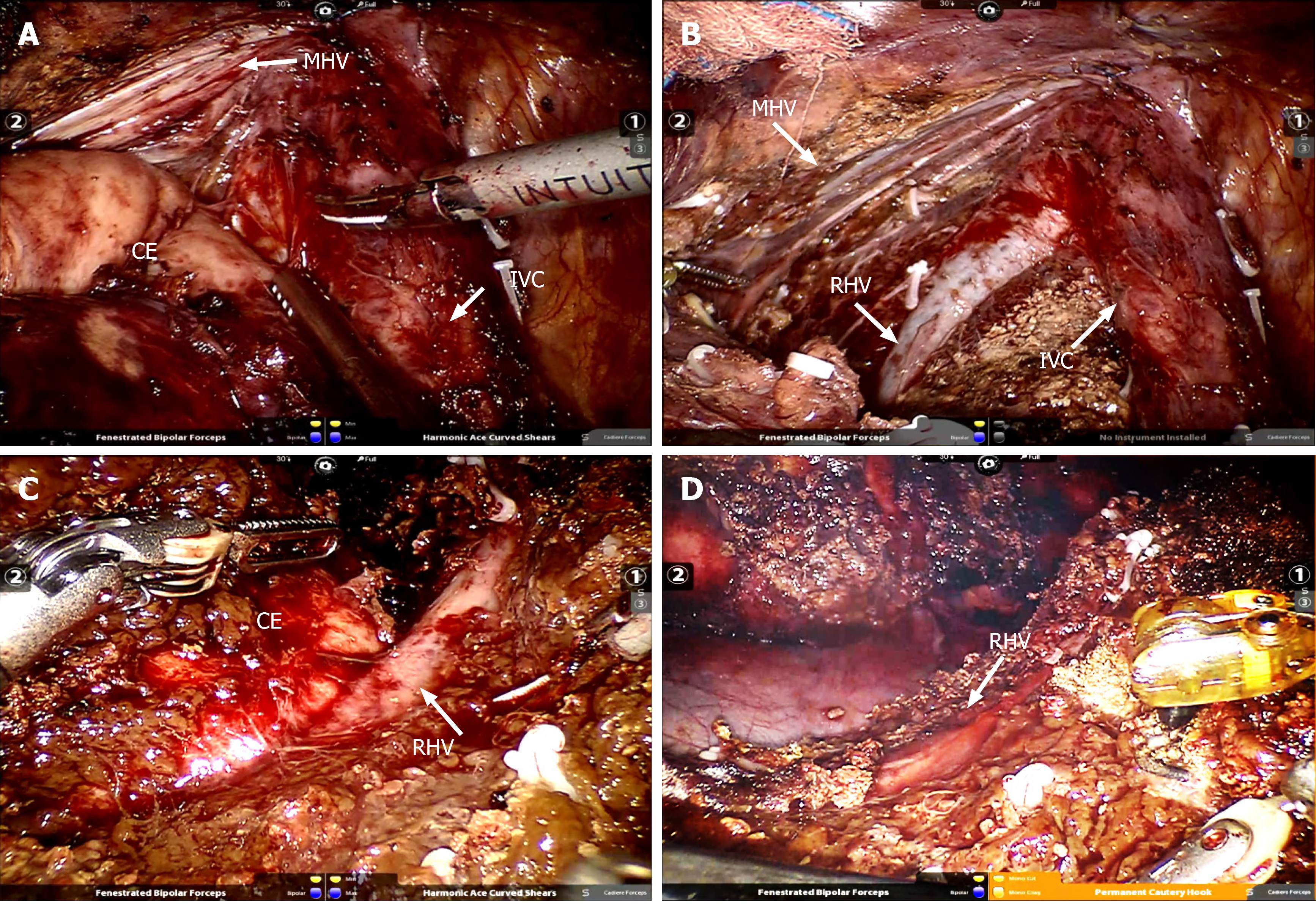
Figure 2 Intraoperative visual field and status after removal of the specimen.
A: Cystic echinococcosis was located in caudate lobe adjacent to the inferior vena and hepatic vein; B: Complete caudate resection; C: cystic echinococcosis was located in segment VII adjacent to the right hepatic vein; D: Segment VII resection. CE: Cystic echinococcosis; IVC: Inferior vena cava; RHV: Right hepatic vein; MHV: Middle hepatic vein.
- Citation: Zhao ZM, Yin ZZ, Meng Y, Jiang N, Ma ZG, Pan LC, Tan XL, Chen X, Liu R. Successful robotic radical resection of hepatic echinococcosis located in posterosuperior liver segments. World J Gastroenterol 2020; 26(21): 2831-2838
- URL: https://www.wjgnet.com/1007-9327/full/v26/i21/2831.htm
- DOI: https://dx.doi.org/10.3748/wjg.v26.i21.2831


