Copyright
©The Author(s) 2019.
World J Gastroenterol. Jan 7, 2019; 25(1): 85-94
Published online Jan 7, 2019. doi: 10.3748/wjg.v25.i1.85
Published online Jan 7, 2019. doi: 10.3748/wjg.v25.i1.85
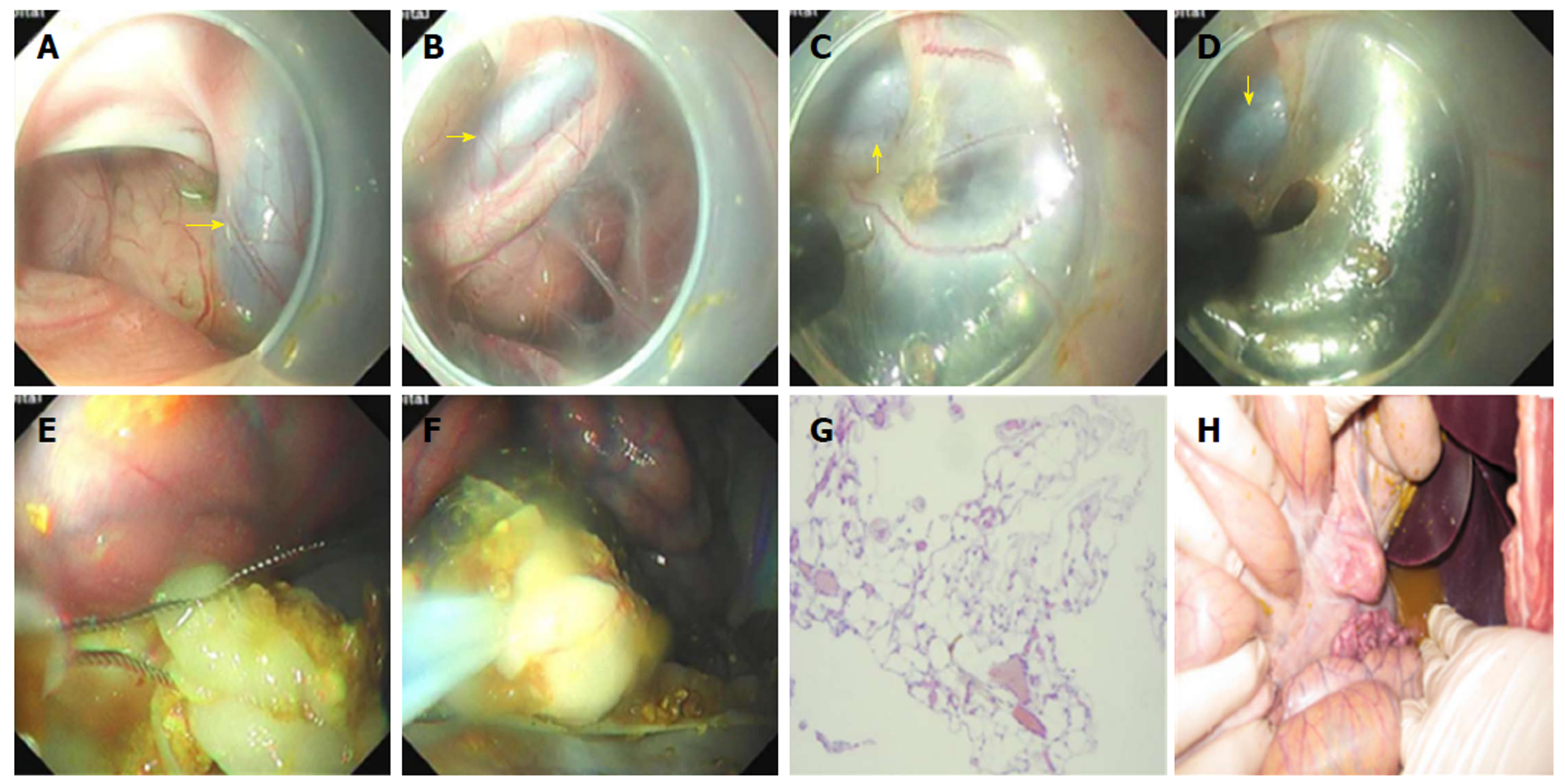
Figure 4 Surgery of retroperitoneal tissue resection.
A: The aorta ventralis was observed in the endoscopic direct status; B: The retroperitoneal space was visible near the aorta ventralis; C and D: Partial resection of tissues in the retroperitoneal space using an electric knife; E and F: Partial resection of the omentum majus using an endoloop; G: Omentum majus tissue stained with hematoxylin and eosin (200 ×); H: A large amount of liquid and chyme in the abdominal cavity.
- Citation: Xiong Y, Chen QQ, Chai NL, Jiao SC, Ling Hu EQ. Endoscopic trans-esophageal submucosal tunneling surgery: A new therapeutic approach for diseases located around the aorta ventralis. World J Gastroenterol 2019; 25(1): 85-94
- URL: https://www.wjgnet.com/1007-9327/full/v25/i1/85.htm
- DOI: https://dx.doi.org/10.3748/wjg.v25.i1.85


