Published online Feb 21, 2017. doi: 10.3748/wjg.v23.i7.1171
Peer-review started: September 8, 2016
First decision: October 20, 2016
Revised: November 9, 2016
Accepted: December 16, 2016
Article in press: December 19, 2016
Published online: February 21, 2017
Processing time: 167 Days and 7.2 Hours
To identify potential anti-cancer constituents in natural extracts that inhibit cancer cell growth and migration.
Our experiments used high dose thymoquinone (TQ) as an inhibitor to arrest LoVo (a human colon adenocarcinoma cell line) cancer cell growth, which was detected by cell proliferation assay and immunoblotting assay. Low dose TQ did not significantly reduce LoVo cancer cell growth. Cyclooxygenase 2 (COX-2) is an enzyme that is involved in the conversion of arachidonic acid into prostaglandin E2 (PGE2) in humans. PGE2 can promote COX-2 protein expression and tumor cell proliferation and was used as a control.
Our results showed that 20 μmol/L TQ significantly reduced human LoVo colon cancer cell proliferation. TQ treatment reduced the levels of p-PI3K, p-Akt, p-GSK3β, and β-catenin and thereby inhibited the downstream COX-2 expression. Results also showed that the reduction in COX-2 expression resulted in a reduction in PGE2 levels and the suppression of EP2 and EP4 activation. Further analysis showed that TG treatment inhibited the nuclear translocation of β-catenin in LoVo cancer cells. The levels of the cofactors LEF-1 and TCF-4 were also decreased in the nucleus following TQ treatment in a dose-dependent manner. Treatment with low dose TQ inhibited the COX-2 expression at the transcriptional level and the regulation of COX-2 expression efficiently reduced LoVo cell migration. The results were further verified in vivo by confirming the effects of TQ and/or PGE2 using tumor xenografts in nude mice.
TQ inhibits LoVo cancer cell growth and migration, and this result highlights the therapeutic advantage of using TQ in combination therapy against colorectal cancer.
Core tip: Prostaglandin E2 (PGE2) induces migration of human LoVo colon cancer cells, and the major mechanism involves the activation of the p-Akt/p-PI3K/p-GSK3β/β-catenin/LEF-1/TCF-4 pathway that ultimately up-regulates cyclooxygenase 2 (COX-2) expression. Thymoquinone (TQ) suppresses cancer cell migration and represents a potential therapeutic target for colon adenocarcinoma metastasis. PGE2 activation of COX-2 and β-catenin to induce human LoVo colon cancer cell migration was blocked by TQ. Our study used cell proliferation assay, immunoblotting assay, immunofluorescence assay, nuclear extraction and in vivo experiments to examine the COX2 protein, which affects the metastasis of highly metastatic LoVo cancer cells treated with TQ.
- Citation: Hsu HH, Chen MC, Day CH, Lin YM, Li SY, Tu CC, Padma VV, Shih HN, Kuo WW, Huang CY. Thymoquinone suppresses migration of LoVo human colon cancer cells by reducing prostaglandin E2 induced COX-2 activation. World J Gastroenterol 2017; 23(7): 1171-1179
- URL: https://www.wjgnet.com/1007-9327/full/v23/i7/1171.htm
- DOI: https://dx.doi.org/10.3748/wjg.v23.i7.1171
Colorectal cancer is one of the most universally diagnosed gastrointestinal cancers and among the main causes of cancer-related death in western developed countries[1,2]. Despite advanced chemotherapeutic treatments, more than 130000 new cases of colon cancer are diagnosed each year[3], causing more than 56000 deaths/year in America[4]. Thymoquinone (TQ) is a phytochemical compound isolated from Nigella sativa that possesses anti-carcinogenic activity and induces apoptosis in tumor cells, and it can interfere with cancer cell survival through different mechanisms[5,6]. Available treatments for cancer include surgical removal, chemotherapy and adjuvant chemotherapy for patients who are strong enough to undergo it. To date, the surgical removal of cancer tissue is considered the most appropriate way to address colon cancer. Our present study investigated the use of phytochemical drugs as a supplementary chemotherapy approach. Laboratory studies have shown that TQ significantly inhibits oral cancer through the p38β MAPK family[7]. Among the hereditary colon cancers, hereditary non-polyposis colon cancer (HNPCC) patients present a particularly high risk for synchronous metastasis via the lymphatic system[8,9]. In this study, we used the colorectal cancer cell line LoVo, which was developed from a 56-year-old colon cancer patient. Many previous studies have verified that prostaglandin E2 (PGE2) promotes cancer development and have considered it a cancer marker; therefore, we used PGE2 as a control[10-12]. PGE2 seems to assist cell survival in colorectal cancer cells by augmenting Ras-MAPK signaling[13].
Compared to normal intestinal tissues, COX-2 expression is 80%-90% higher in colorectal cancers. Cancers of the head, breast, cervix, bladder and gastrointestinal system have also shown high levels of COX-2 expression[14-16]. COX-2/PGE2 signaling affects cell physiology in multiple tumor types and maintains colorectal tumorigenesis[12,17]. PGE2 as a proangiogenic factor is associated with transformed vascular permeability and angiogenesis[18]. COX-2 expression is thought to contribute to the principal PGE2 metabolic product[19,20]. Some non-steroidal anti-inflammatory drugs (NSAIDs) and vegetables produce anti-tumor effects that reduce PGE2 synthesis or inhibit COX-2[21-24]. In our experiments, we sought to identify compounds similar to NSAIDs or adjunct drugs to increase the effectiveness of cancer chemotherapy. Our experimental drug, TQ, has promising anti-tumor effects, and it inhibited the incidence of fore-stomach tumors and fibrosarcoma tumors and increased cellular longevity[25,26]. We previously evaluated PGE2-induced migration in human LoVo cancer cells, and the major mechanism involves the activation of the p-Akt/p-PI3K/p-GSK3β/β-catenin pathway that ultimately up-regulates COX-2 expression (unpublished data). After the addition of TQ, the exact anticancer mechanism produced by PGE2 was determined. Previous studies have demonstrated that β-catenin translocation, which includes co-interaction with and activation of the promoters LEF-1 and TCF-4, subsequently modulates downstream gene expression[27]. The nuclear cofactors LEF and TCF were triggered to initiate the transcription and translation of COX-2[28]. Cell metastasis efficiency is a focus of our work because it correlates with COX-2 activity[29,30]. Moreover, cell migration is promoted due to COX-2 expression[31]. Numerous studies of animals treated with TQ have demonstrated that TQ is not toxic[32-34].
Our study used immunoblotting assays, immunofluorescence assays, nuclear extraction and in vivo experiments to examine the COX2 protein, which affects the metastasis of highly metastatic LoVo cancer cells treated with TQ.
The human colon cancer cell line LoVo was obtained from the American Tissue Culture Collection (ATCC) (Rockville, MD, United States). LoVo cells were established from metastatic nodules that were resected from a 56-year-old colon adenocarcinoma patient.
We utilized antibodies against the following proteins: phospho-PI3K, phospho-Akt, COX-2, phospho-GSK3β, β-catenin, LEF-1, HADAC-1 (Santa Cruz Biotechnology, Inc. Santa Cruz, CA, United States), and TCF-4 (Cell Signaling Technology, Inc. Beverly, MA, United States). α-tubulin and β-actin (Santa Cruz Biotechnology, Inc. Santa Cruz, CA, United States) were used as loading controls. The following horseradish peroxidase-conjugated antibodies were purchased from Santa Cruz Biotechnology, Inc. (CA, United States): goat anti-mouse IgG, goat anti-rabbit IgG, and rabbit anti-goat IgG. Nude mice were purchased from the National Laboratory Animal Center (NLAC).
The colon cancer cell line LoVo was cultured in 10-cm2 culture dishes in Dulbecco’s modified Eagle’s medium (DMEM) supplemented with 100 μg/mL penicillin, 100 μg/mL streptomycin, 2 mmol/L glutamine, 1 mmol/L HEPES buffer, and 10% fetal bovine serum (FBS) in humidified air (5% CO2) at 37 °C.
LoVo cells were seeded at a density of 1.5 × 10⁴ cells per well in 24-well plates, and after 24 h the cells were treated with different concentrations of TQ varying from 0 to 20 μmol/L (Sigma Aldrich, St Louis MO, United States) dissolved in DMSO. An MTT (3-(4,5-Dimethylthiazol-2-yl)-2,5-diphenyltetrazolium bromide) assay was used to determine living cells 24 h after treatment. Culture supernatants were removed, and MTT (Sigma, United States) in phosphate-buffered saline (PBS, pH 7.4) was added to each well. After 4 h of incubation at 37 °C, the MTT solution was removed, and DMSO was added to dissolve the resultant formazan crystals. Absorbance was read at 540 nm in a Flexstation 3 device (MDS Analytical Technologies, Canada). The percentage viability was calculated as (test-background)/(control-background) × 100.
Cultured LoVo cells were washed with cold PBS and resuspended in lysis buffer [50 mmol/L Tris (pH 7.5), 0.5 mol/L NaCl, 1.0 mmol/L EDTA (pH 7.5), 10% glycerol, 1 mmol/L BME, 1% IGEPAL-630, and a proteinase inhibitor cocktail (Roche Molecular Biochemical)] to isolate total proteins. After incubation for 30 min on ice, the supernatant was collected by centrifugation at 12000 ×g for 15 min at 4 °C. Protein concentration was then determined using the Bradford method. Samples containing equal protein amounts (60 μg) were loaded and analyzed using immunoblotting analysis. Proteins were separated using 10% SDS-PAGE and transferred onto PVDF membranes (Millipore, Belford, MA, United States). The membranes were blocked with blocking buffer (5% non-fat dry milk, 20 mmol/L Tris-HCl, pH 7.6, 150 mmol/L NaCl, and 0.1% Tween 20) for at least 1 h at room temperature. The membranes were incubated with primary antibodies in the above solution on an orbital shaker at 4 °C overnight. Following the primary antibody incubations, the membranes were incubated with horseradish peroxidase-linked secondary antibodies (anti-rabbit, anti-mouse, or anti-goat IgG). Detection was performed using ECL reagents and a digital imaging system.
Cytoplasmic and nuclear fractions were obtained using extraction reagents containing membrane lysis buffer [10 mmol/L HEPES (pH 8.0), 1.5 mmol/L MgCl2, 10 mmol/L KCl, 1 mmol/L DDT, and proteinase inhibitor] and nuclear lysis buffer [20 mmol/L HEPES (pH 8.0), 1.5 mmol/L MgCl2, 10 mmol/L NaCl, 1 mmol/L DDT, 0.2 mmol/L EDTA, 0.25 mol/L glycerol, and proteinase inhibitor]. Following treatment, the cells were resuspended in PBS, incubated with ice-cold membrane lysis buffer for 10 min, and centrifuged at 12000 rpm for 2 min to pellet nuclei. The supernatant was stored for use as a cytoplasmic fraction, and the nuclear pellet was lysed with nuclear lysis buffer to obtain a nuclear fraction.
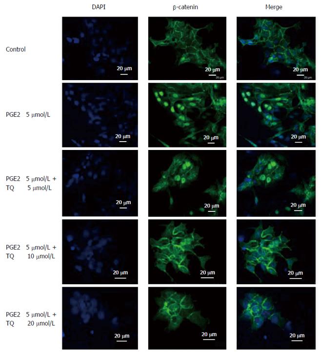
LoVo cells were treated with increasing TQ dosages (5, 10, and 20 μmol/L) for 24 h, and PGE2 (5 μmol/L) was added for the last six hours. An immunofluorescence assay was performed on the LoVo cells using a β-catenin antibody (1:250) directed against a target of interest. A fluorophore-conjugated secondary antibody (green) that was directed against the primary antibody was used for detection, and a blue fluorescent DAPI nuclear acid counterstain that preferentially stains dsDNA was included. Anti-β-catenin (green) merged with DAPI (blue) is also shown in the resultant confocal microscopy images.
A migration assay was performed using a 48-well Boyden chamber (Neuro Probe) plate with 8-μm pore size polycarbonate membrane filters[35]. The lower compartment was filled with DMEM containing 10% FBS. LoVo cells were placed in the upper part of the Boyden chamber, which contained serum-free medium, and incubated for 4-8 h. After incubation, cells on the membrane filter were fixed with methanol and stained with 0.05% Giemsa for 1 h. The cells on the upper filter surface were removed with a cotton swab. The filters were then rinsed in double-distilled water for additional stain leaching. The cells were then air-dried for 20 min. Migratory phenotypes were determined by counting the cells that migrated to the lower side of the filter using microscopy at 200 × and 400 × magnification. Ten fields were blindly selected and counted as the mean cell number in each filter. The sample was assayed in triplicate.
Twelve nude mice were bred in the animal care facility of the Chinese Medicine Library (Taichung, Taiwan) and were purchased from The National Laboratory Animal Center. The animals received subcutaneous injections of LoVo colorectal cancer cells (1 × 10⁶ cells per injection) in the back. Ambient temperature was maintained at 25 °C, and the mice were kept on an artificial 12-h light-dark cycle, with the light period beginning at 7:00 am. All of the protocols were approved by the Institutional Animal Care and Use Committee of China Medical University, Taichung, Taiwan, ROC.
LoVo cells were treated with PGE2 (5 μmol/L) for 24 h and were then injected subcutaneously into the mice (1 × 10⁶ cells). After one week, various concentrations (0.5, 10 and 20 μmol/L) of TQ were administered for three weeks (3 times per week) by intraperitoneal injection. All protocols were reviewed and approved by the Institutional Review Board (IRB, ethical clearance number 104-223-N), Animal Care and Use Committee of China Medical University, Taichung, Republic of China, and the study was conducted in accordance with the principles of laboratory animal care[36].
Each experiment was duplicated at least three times. The results are presented as the mean ± SE, and statistical comparisons were made using Student’s t-test. Significance was defined at P < 0.05 or 0.01.
We first tested the effect of TQ on the viability of LoVo cells. Several TQ concentrations were used to evaluate cell viability after co-culture for 24 h. The results indicated that 20 μmol/L TQ can significantly reduce LoVo cell proliferation (Figure 1).
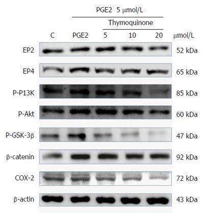
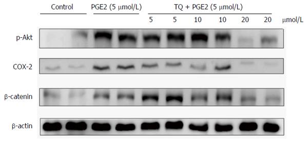
Previous reports have demonstrated that PGE2 increases COX-2 expression via activation of the EP4/β-catenin pathway, suggesting that PGE2 regulates p-PI3K, p-AKT, and p-GSK-3β expression in LoVo cells. PGE2 has been reported to activate downstream signaling via EP2 and EP4 to elicit various biological responses. As shown in Figure 2, TQ treatment reduced the up-regulation of COX-2 expression by PGE2, and β-catenin had an obvious effect on EP2 and EP4 regulation by PGE2.
β-catenin is a key protein that affects nuclear COX-2 transcription. We used an immunofluorescence assay to evaluate β-catenin protein levels in the cytosol and nucleus. Confocal microscopy confirmed that TQ treatment led to the translocation of β-catenin into the LoVo cell nucleus (Figure 3).
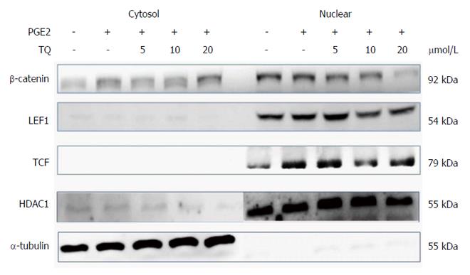
Previous studies have demonstrated that the translocation of β-catenin involves co-interaction with and activation of the promoters LEF-1 and TCF-4, which subsequently modulate downstream gene expression. Evaluation of isolated LoVo cell nuclei showed that β-catenin translocation decreased in the nucleus, but its interactions with the cofactors LEF-1 and TCF-4 did not change. We observed a concentration-dependent decrease in the levels of β-catenin and the proteins LEF-1 and TCF-4 in the nucleus following TQ treatment (Figure 4).
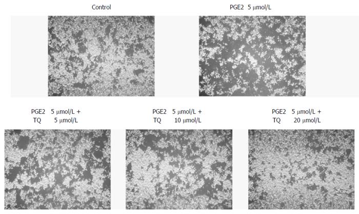
Migration is an important step in LoVo colon cancer progression because this cancer metastasizes supraclavicular lymph nodes. We performed a Boyden chamber migration assay to determine the effect of TQ on LoVo cells and found that TQ inhibited LoVo cell migration in a dose-dependent manner (Figure 5).
To verify previous results on the growth inhibition and anti-metastatic effects of TQ in human LoVo cells, the therapeutic potential of TQ in a pre-subcutaneous LoVo cell model in nude mice was determined. TQ was administered for 30 consecutive days. The drug did not induce death or weight loss of more than 15% to 20% of original weight, which is a very sensitive parameter for perniciousness in mice. The effects of TQ on PGE2-associated protein markers for inflammation and metastasis in the nude mouse xenograft model were then determined. COX-2 and β-catenin protein levels in different tissue groups in the nude mice were down-regulated by TQ treatment in a concentration-dependent manner (Figure 6).
TQ was shown to be an anticancer agent by virtue of its anti-proliferative potential and capacity to induce cell cycle arrest[37]. In our previous studies, COX-2 protein was found to be regulated by PGE2, resulting in cell proliferation and migration. Here, a 20 μmol/L concentration of TQ, which is 50% of the non-cytotoxic concentration determined by MTT assay, arrested the migration of LoVo colorectal cancer cells. We tested TQ on LoVo colorectal cancer cells that were already induced by PGE2 (5 μmol/L); these cells exhibited significantly enhanced performance over the control group. Our experimental drug TQ inhibited the survival pathway proteins p-PI3K, p-Akt, p-GSK3β, β-catenin and COX-2.
When β-catenin is translocated from the cytosol into the nucleus, it plays a role in the transcription and translation of COX-2[28]. The translocation of β-catenin from the cytosol into the nucleus also plays a role in the transcription and translation of COX-2.
Nuclear isolation techniques and immunofluorescence were used to determine the localization of β-catenin and the COX-2 transcription cofactors LEF-1 and TCF-4[38]. Our experiments showed that TQ suppresses β-catenin translocation induced by PGE2, and the expression of both LEF-1 and TCF-4 was also suppressed. This result suggests that β-catenin loses its ability to translocate into the nucleus and bind to LEF-1 and TCF-4 following TQ treatment. These cofactors were also affected by TQ with a gradually declining, concentration-dependent trend. Immunofluorescence confocal microscopy showed fluorescent images of anti-β-catenin (green) and DAPI staining (blue) in the panel representing the cell nucleus. These results indicate that TQ treatment inhibited β-catenin translocation into the nucleus and subsequently reduced COX-2 activation.
We can observe the impact of TQ on COX-2 by assessing these translation- and transcription-associated proteins. COX-2 plays a central role in cell migration. These results showed that decreasing COX-2 levels will correspondingly reduce cell migration. In this context, we demonstrated that TQ significantly reduces metastasis in vivo. However, we investigated the therapeutic potential of TQ using a highly aggressive human LOVO cancer cell xenograft nude mouse model. p-Akt, β-catenin and COX-2 expression substantially decreased. TQ inhibited LoVo colorectal cancer cell growth and inhibited migration. In the future, we will evaluate the therapeutic advantage of combining chemotherapeutic agents for colorectal cancer.
Several types of phytochemicals are used for cancer chemotherapy. The aim was to identify potential anti-cancer constituents in natural extracts that inhibit cancer cell growth and migration. Thymoquinone (TQ) is a phytochemical compound isolated from Nigella sativa. Previous data show that TQ suppresses the activation of AKT, inhibits cellular proliferation, and shows anti-oxidant/anti-inflammatory effects.
Prostaglandin E2 (PGE2) induces migration of human LoVo colon cancer cells, and the major mechanism involves the activation of the p-Akt/p-PI3K/p-GSK3β/β-catenin/LEF-1/TCF-4 pathway that ultimately up-regulates cyclooxygenase 2 (COX-2) expression.
TQ suppresses cancer cell migration and represents a potential therapeutic target for colon adenocarcinoma metastasis. PGE2 activation of COX-2 and β-catenin to induce human LoVo colon cancer cell migration was blocked by thymoquinone.
The results reveal that TQ can be considered a potential treatment strategy for advanced stage colon cancer treatment.
The study described in this paper investigated the use of phytochemical drugs as a supplementary chemotherapy approach. For this purpose the authors used the high dose of TQ as an inhibitor to arrest LoVo (a human colon adenocarcinoma cell line) cell growth. The authors used immunoblotting assays, immunofluorescence assays, nuclear extraction and in vivo experiments to examine the COX2 protein, which affects the metastasis of highly metastatic LoVo cancer cells treated with TQ.
Manuscript source: Unsolicited manuscript
Specialty type: Gastroenterology and hepatology
Country of origin: Taiwan
Peer-review report classification
Grade A (Excellent): 0
Grade B (Very good): B
Grade C (Good): 0
Grade D (Fair): 0
Grade E (Poor): 0
P- Reviewer: Kunac N S- Editor: Gong ZM L- Editor: Wang TQ E- Editor: Wang CH
| 1. | Gali-Muhtasib H, Ocker M, Kuester D, Krueger S, El-Hajj Z, Diestel A, Evert M, El-Najjar N, Peters B, Jurjus A. Thymoquinone reduces mouse colon tumor cell invasion and inhibits tumor growth in murine colon cancer models. J Cell Mol Med. 2008;12:330-342. [RCA] [PubMed] [DOI] [Full Text] [Full Text (PDF)] [Cited by in Crossref: 134] [Cited by in RCA: 113] [Article Influence: 6.6] [Reference Citation Analysis (0)] |
| 2. | Lin TY, Fan CW, Maa MC, Leu TH. Lipopolysaccharide-promoted proliferation of Caco-2 cells is mediated by c-Src induction and ERK activation. Biomedicine (Taipei). 2015;5:5. [RCA] [PubMed] [DOI] [Full Text] [Full Text (PDF)] [Cited by in Crossref: 34] [Cited by in RCA: 55] [Article Influence: 5.5] [Reference Citation Analysis (0)] |
| 3. | Jemal A, Thomas A, Murray T, Thun M. Cancer statistics, 2002. CA Cancer J Clin. 2002;52:23-47. [PubMed] |
| 4. | Jemal A, Tiwari RC, Murray T, Ghafoor A, Samuels A, Ward E, Feuer EJ, Thun MJ. Cancer statistics, 2004. CA Cancer J Clin. 2004;54:8-29. [PubMed] |
| 5. | Woo CC, Kumar AP, Sethi G, Tan KH. Thymoquinone: potential cure for inflammatory disorders and cancer. Biochem Pharmacol. 2012;83:443-451. [RCA] [PubMed] [DOI] [Full Text] [Cited by in Crossref: 304] [Cited by in RCA: 328] [Article Influence: 23.4] [Reference Citation Analysis (0)] |
| 6. | Towhid ST, Schmidt EM, Schmid E, Münzer P, Qadri SM, Borst O, Lang F. Thymoquinone-induced platelet apoptosis. J Cell Biochem. 2011;112:3112-3121. [RCA] [PubMed] [DOI] [Full Text] [Cited by in Crossref: 13] [Cited by in RCA: 15] [Article Influence: 1.2] [Reference Citation Analysis (0)] |
| 7. | Abdelfadil E, Cheng YH, Bau DT, Ting WJ, Chen LM, Hsu HH, Lin YM, Chen RJ, Tsai FJ, Tsai CH. Thymoquinone induces apoptosis in oral cancer cells through p38β inhibition. Am J Chin Med. 2013;41:683-696. [RCA] [PubMed] [DOI] [Full Text] [Cited by in Crossref: 44] [Cited by in RCA: 47] [Article Influence: 3.9] [Reference Citation Analysis (0)] |
| 8. | Kalady MF. Surgical management of hereditary nonpolyposis colorectal cancer. Adv Surg. 2011;45:265-274. [PubMed] |
| 9. | Wu Z, Zhang S, Aung LH, Ouyang J, Wei L. Lymph node harvested in laparoscopic versus open colorectal cancer approaches: a meta-analysis. Surg Laparosc Endosc Percutan Tech. 2012;22:5-11. [RCA] [PubMed] [DOI] [Full Text] [Cited by in Crossref: 29] [Cited by in RCA: 30] [Article Influence: 2.3] [Reference Citation Analysis (0)] |
| 10. | Chang SH, Liu CH, Conway R, Han DK, Nithipatikom K, Trifan OC, Lane TF, Hla T. Role of prostaglandin E2-dependent angiogenic switch in cyclooxygenase 2-induced breast cancer progression. Proc Natl Acad Sci USA. 2004;101:591-596. [RCA] [PubMed] [DOI] [Full Text] [Cited by in Crossref: 286] [Cited by in RCA: 304] [Article Influence: 13.8] [Reference Citation Analysis (0)] |
| 11. | Eruslanov E, Kaliberov S, Daurkin I, Kaliberova L, Buchsbaum D, Vieweg J, Kusmartsev S. Altered expression of 15-hydroxyprostaglandin dehydrogenase in tumor-infiltrated CD11b myeloid cells: a mechanism for immune evasion in cancer. J Immunol. 2009;182:7548-7557. [RCA] [PubMed] [DOI] [Full Text] [Cited by in Crossref: 57] [Cited by in RCA: 65] [Article Influence: 4.1] [Reference Citation Analysis (0)] |
| 12. | Greenhough A, Smartt HJ, Moore AE, Roberts HR, Williams AC, Paraskeva C, Kaidi A. The COX-2/PGE2 pathway: key roles in the hallmarks of cancer and adaptation to the tumour microenvironment. Carcinogenesis. 2009;30:377-386. [RCA] [PubMed] [DOI] [Full Text] [Cited by in Crossref: 848] [Cited by in RCA: 937] [Article Influence: 58.6] [Reference Citation Analysis (0)] |
| 13. | Kaidi A, Qualtrough D, Williams AC, Paraskeva C. Direct transcriptional up-regulation of cyclooxygenase-2 by hypoxia-inducible factor (HIF)-1 promotes colorectal tumor cell survival and enhances HIF-1 transcriptional activity during hypoxia. Cancer Res. 2006;66:6683-6691. [RCA] [PubMed] [DOI] [Full Text] [Cited by in Crossref: 226] [Cited by in RCA: 236] [Article Influence: 12.4] [Reference Citation Analysis (0)] |
| 14. | Voutsadakis IA. Pathogenesis of colorectal carcinoma and therapeutic implications: the roles of the ubiquitin-proteasome system and Cox-2. J Cell Mol Med. 2007;11:252-285. [RCA] [PubMed] [DOI] [Full Text] [Full Text (PDF)] [Cited by in Crossref: 53] [Cited by in RCA: 61] [Article Influence: 3.4] [Reference Citation Analysis (0)] |
| 15. | Sano H, Kawahito Y, Wilder RL, Hashiramoto A, Mukai S, Asai K, Kimura S, Kato H, Kondo M, Hla T. Expression of cyclooxygenase-1 and -2 in human colorectal cancer. Cancer Res. 1995;55:3785-3789. [PubMed] |
| 16. | Eberhart CE, Coffey RJ, Radhika A, Giardiello FM, Ferrenbach S, DuBois RN. Up-regulation of cyclooxygenase 2 gene expression in human colorectal adenomas and adenocarcinomas. Gastroenterology. 1994;107:1183-1188. [PubMed] |
| 17. | Brown JR, DuBois RN. COX-2: a molecular target for colorectal cancer prevention. J Clin Oncol. 2005;23:2840-2855. [RCA] [PubMed] [DOI] [Full Text] [Cited by in Crossref: 387] [Cited by in RCA: 413] [Article Influence: 20.7] [Reference Citation Analysis (0)] |
| 18. | Form DM, Auerbach R. PGE2 and angiogenesis. Proc Soc Exp Biol Med. 1983;172:214-218. [PubMed] |
| 19. | Pugh S, Thomas GA. Patients with adenomatous polyps and carcinomas have increased colonic mucosal prostaglandin E2. Gut. 1994;35:675-678. [PubMed] |
| 20. | Rigas B, Goldman IS, Levine L. Altered eicosanoid levels in human colon cancer. J Lab Clin Med. 1993;122:518-523. [PubMed] |
| 21. | Hanif R, Pittas A, Feng Y, Koutsos MI, Qiao L, Staiano-Coico L, Shiff SI, Rigas B. Effects of nonsteroidal anti-inflammatory drugs on proliferation and on induction of apoptosis in colon cancer cells by a prostaglandin-independent pathway. Biochem Pharmacol. 1996;52:237-245. [PubMed] |
| 22. | Elder DJ, Halton DE, Hague A, Paraskeva C. Induction of apoptotic cell death in human colorectal carcinoma cell lines by a cyclooxygenase-2 (COX-2)-selective nonsteroidal anti-inflammatory drug: independence from COX-2 protein expression. Clin Cancer Res. 1997;3:1679-1683. [PubMed] |
| 23. | Chan TA, Morin PJ, Vogelstein B, Kinzler KW. Mechanisms underlying nonsteroidal antiinflammatory drug-mediated apoptosis. Proc Natl Acad Sci USA. 1998;95:681-686. [PubMed] |
| 24. | Ciou SY, Hsu CC, Kuo YH, Chao CY. Effect of wild bitter gourd treatment on inflammatory responses in BALB/c mice with sepsis. Biomedicine (Taipei). 2014;4:17. [RCA] [PubMed] [DOI] [Full Text] [Full Text (PDF)] [Cited by in Crossref: 23] [Cited by in RCA: 23] [Article Influence: 2.1] [Reference Citation Analysis (0)] |
| 25. | Badary OA, Gamal El-Din AM. Inhibitory effects of thymoquinone against 20-methylcholanthrene-induced fibrosarcoma tumorigenesis. Cancer Detect Prev. 2001;25:362-368. [PubMed] |
| 26. | Badary OA, Al-Shabanah OA, Nagi MN, Al-Rikabi AC, Elmazar MM. Inhibition of benzo(a)pyrene-induced forestomach carcinogenesis in mice by thymoquinone. Eur J Cancer Prev. 1999;8:435-440. [PubMed] |
| 27. | Kriegl L, Horst D, Reiche JA, Engel J, Kirchner T, Jung A. LEF-1 and TCF4 expression correlate inversely with survival in colorectal cancer. J Transl Med. 2010;8:123. [RCA] [PubMed] [DOI] [Full Text] [Full Text (PDF)] [Cited by in Crossref: 46] [Cited by in RCA: 47] [Article Influence: 3.1] [Reference Citation Analysis (0)] |
| 28. | Nuñez F, Bravo S, Cruzat F, Montecino M, De Ferrari GV. Wnt/β-catenin signaling enhances cyclooxygenase-2 (COX2) transcriptional activity in gastric cancer cells. PLoS One. 2011;6:e18562. [RCA] [PubMed] [DOI] [Full Text] [Full Text (PDF)] [Cited by in Crossref: 80] [Cited by in RCA: 91] [Article Influence: 6.5] [Reference Citation Analysis (0)] |
| 29. | Xu L, Stevens J, Hilton MB, Seaman S, Conrads TP, Veenstra TD, Logsdon D, Morris H, Swing DA, Patel NL. COX-2 inhibition potentiates antiangiogenic cancer therapy and prevents metastasis in preclinical models. Sci Transl Med. 2014;6:242ra84. [RCA] [PubMed] [DOI] [Full Text] [Cited by in Crossref: 158] [Cited by in RCA: 157] [Article Influence: 14.3] [Reference Citation Analysis (0)] |
| 30. | Emenaker NJ, Zudaire E, St Croix B. Chemoprevention of metastasis. Oncotarget. 2014;5:6556-6557. [PubMed] |
| 31. | Zhao L, Wu Y, Xu Z, Wang H, Zhao Z, Li Y, Yang P, Wei X. Involvement of COX-2/PGE2 signalling in hypoxia-induced angiogenic response in endothelial cells. J Cell Mol Med. 2012;16:1840-1855. [RCA] [PubMed] [DOI] [Full Text] [Full Text (PDF)] [Cited by in Crossref: 35] [Cited by in RCA: 44] [Article Influence: 3.4] [Reference Citation Analysis (0)] |
| 32. | Kirui PK, Cameron J, Benghuzzi HA, Tucci M, Patel R, Adah F, Russell G. Effects of sustained delivery of thymoqiunone on bone healing of male rats. Biomed Sci Instrum. 2004;40:111-116. [PubMed] |
| 33. | Hosseinzadeh H, Parvardeh S. Anticonvulsant effects of thymoquinone, the major constituent of Nigella sativa seeds, in mice. Phytomedicine. 2004;11:56-64. [RCA] [PubMed] [DOI] [Full Text] [Cited by in Crossref: 137] [Cited by in RCA: 132] [Article Influence: 6.3] [Reference Citation Analysis (0)] |
| 34. | Attoub S, Sperandio O, Raza H, Arafat K, Al-Salam S, Al Sultan MA, Al Safi M, Takahashi T, Adem A. Thymoquinone as an anticancer agent: evidence from inhibition of cancer cells viability and invasion in vitro and tumor growth in vivo. Fundam Clin Pharmacol. 2013;27:557-569. [RCA] [PubMed] [DOI] [Full Text] [Cited by in Crossref: 86] [Cited by in RCA: 93] [Article Influence: 7.2] [Reference Citation Analysis (0)] |
| 35. | Hsieh YC, Hsieh SJ, Chang YS, Hsueh CM, Hsu SL. The lipoxygenase inhibitor, baicalein, modulates cell adhesion and migration by up-regulation of integrins and vinculin in rat heart endothelial cells. Br J Pharmacol. 2007;151:1235-1245. [RCA] [PubMed] [DOI] [Full Text] [Cited by in Crossref: 14] [Cited by in RCA: 15] [Article Influence: 0.8] [Reference Citation Analysis (0)] |
| 36. | U.S. National Institutes of Health. Laboratory animal welfare; proposed U.S. government principles for the utilization and care of vertebrate animals used in testing, research and training. Fed Regist. 1984;49:29350-29351. [PubMed] |
| 37. | Jrah-Harzallah H, Ben-Hadj-Khalifa S, Almawi WY, Maaloul A, Houas Z, Mahjoub T. Effect of thymoquinone on 1,2-dimethyl-hydrazine-induced oxidative stress during initiation and promotion of colon carcinogenesis. Eur J Cancer. 2013;49:1127-1135. [RCA] [PubMed] [DOI] [Full Text] [Cited by in Crossref: 46] [Cited by in RCA: 46] [Article Influence: 3.5] [Reference Citation Analysis (0)] |
| 38. | Daniels DL, Weis WI. Beta-catenin directly displaces Groucho/TLE repressors from Tcf/Lef in Wnt-mediated transcription activation. Nat Struct Mol Biol. 2005;12:364-371. [RCA] [PubMed] [DOI] [Full Text] [Cited by in Crossref: 388] [Cited by in RCA: 431] [Article Influence: 21.6] [Reference Citation Analysis (0)] |


