Copyright
©The Author(s) 2016.
World J Gastroenterol. May 7, 2016; 22(17): 4330-4337
Published online May 7, 2016. doi: 10.3748/wjg.v22.i17.4330
Published online May 7, 2016. doi: 10.3748/wjg.v22.i17.4330
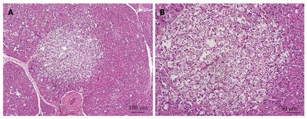
Figure 2 Photomicrographs of the left lobe (tail) of porcine pancreas group Group Papilla.
A: Pancreas showing focal necrosis perivascular with evidence of vacuolization in acinar cells; B: Magnification of previous image, view of acinar cell´s cytoplasm vacuolated. The nuclei are fragmented and shrunken. Hematoxylin and eosin staining.
- Citation: Latorre R, López-Albors O, Soria F, Candanosa E, Pérez-Cuadrado E. Effect of the manipulation of the duodenal papilla during double balloon enteroscopy. World J Gastroenterol 2016; 22(17): 4330-4337
- URL: https://www.wjgnet.com/1007-9327/full/v22/i17/4330.htm
- DOI: https://dx.doi.org/10.3748/wjg.v22.i17.4330


