Published online Mar 7, 2015. doi: 10.3748/wjg.v21.i9.2725
Peer-review started: July 25, 2014
First decision: August 15, 2014
Revised: September 4, 2014
Accepted: December 5, 2014
Article in press: December 8, 2014
Published online: March 7, 2015
Processing time: 227 Days and 21.3 Hours
AIM: To assess the feasibility and safety of the use of soehendra stent retriever as a new technique for biliary access in endoscopic ultrasound-guided biliary drainage.
METHODS: The medical records and endoscopic reports of the patients who underwent endoscopic ultrasound-guided biliary drainage (EUS-BD) owing to failed endoscopic retrograde cholangiopancreatography in our institute between June 2011 and January 2014 were collected and reviewed. All the procedures were performed in the endoscopic suite under intravenous sedation with propofol and full anaesthetic monitoring. Then we used the Soehendra stent retriever as new equipment for neo-tract creation and dilation when performing EUS-BD procedures. The patients were observed in the recovery room for 1-2 h and transferred to the regular ward, patients’ clinical data were reviewed and analysed, clinical outcomes were defined by using several different criteria. Data were analysed by using SPSS 13 and presented as percentages, means, and medians.
RESULTS: A total of 12 patients were enrolled. The most common indications for EUS-BD in this series were failed common bile duct cannulation, duodenal obstruction, failed selective intrahepatic duct cannulation, and surgical altered anatomy for 50%, 25%, 16.7%, and 8.3%, respectively. Seven patients underwent EUS-guided hepaticogastrostomy (58.3%), and 5 underwent EUS-guided choledochoduodenostomy (41.7%). The technical success rate was 100%, while the clinical success rate was 91.7%. Major and minor complications occurred in 16.6% and 33.3% of patients, respectively, but there were no procedure-related death.
CONCLUSION: Soehendra stent retriever could be used as an alternative instrument for biliary access in endoscopic ultrasound guided biliary drainage.
Core tip: Endoscopic ultrasound-guided biliary drainage is considered an alternative treatment for the patients who failed endoscopic retrograde cholangiopancreatography. We proposed that the technical and clinical success rate, including complications were related to the techniques of biliary access which demonstrated an impressive outcomes using the selfmade-tapered tip Teflon catheters for neo-tract creation. In this case series, we would like to shared our experiences using Soehendra stent retrievers as a new technique for biliary access in endoscopic ultrasound-guided biliary drainage.
- Citation: Prachayakul V, Aswakul P. Feasibility and safety of using Soehendra stent retriever as a new technique for biliary access in endoscopic ultrasound-guided biliary drainage. World J Gastroenterol 2015; 21(9): 2725-2730
- URL: https://www.wjgnet.com/1007-9327/full/v21/i9/2725.htm
- DOI: https://dx.doi.org/10.3748/wjg.v21.i9.2725
Endoscopic ultrasound-guided biliary drainage (EUS-BD), both choledochoduodenostomy and hepaticogastrostomy, is considered an alternative treatment for patients with difficult malignant biliary obstruction in whom endoscopic retrograde cholangiopancreatography (ERCP) for palliative biliary drainage failed.
EUS-BD is currently comparative or superior to conventional percutaneous transhepatic biliary drainage in some retrospective studies. This procedure has been reported effective and safe when performed by experts, with 85%-100% success rates and 5%-35% complication rates[1-8]. The important systematic procedures were localization and puncture, followed by guidewire negotiation into the desired position, followed by biloenteric fistula creation and dilatation by using an over-the-guidewire technique, and finally stent placement. However, the important issues of high success and low complication rate of EUS-BD makes it an appropriate technique for biliary access. The techniques for biliary access were classified into neotract creation with cauterized or non-cauterized and neotract dilatation with graded or balloon dilation. Many studies have reported techniques and equipment used in these procedure. The common techniques for biliary access included using a needle knife to create the neotract followed by balloon dilatation with different balloon types. However, the authors usually do not use cauterization and balloon dilation for biliary access in EUS-BD owing to concerns about complications, especially perforation and bile leakage. To date, the author has had more experience with a non-cauterized and graded dilatation technique by using the self-made, tapered-tip, Teflon catheter, which provided high success and low complication rates[9,10]. However, the limitations of self-made, tapered-tip, Teflon catheters for biliary access warrant the use of alternative equipment in some cases. The Soehendra stent retriever was first used for biliary stent retrieval. It was recently proven useful in many therapeutic applications, especially dilatation of a tightly strictured pancreatic duct, bile duct, and puncture hole in patients who underwent EUS-guided cystogastrostomy[11-13]. Therefore, the authors were very grateful to share our experiences about using this well-designed multi-purposes equipment in EUS-BD procedures.
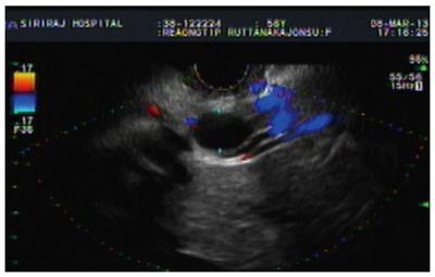
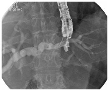
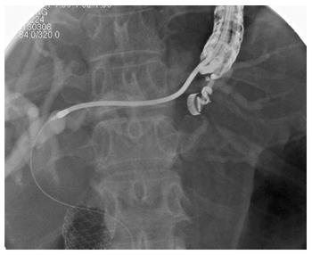
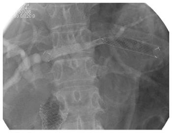
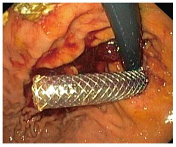
This study was approved by Siriraj Institutional Review Board committee. The medical records and endoscopic reports of the patients who underwent EUS-BD owing to failed ERCP in our institute between June 2011 and January 2014 were collected and reviewed. All patients underwent EUS-BD performed by a single endoscopist (VP) and provided informed consent before the procedures. All the procedures were performed in the endoscopic suite under intravenous sedation with propofol and full anaesthetic monitoring. The patient was in the left lateral decubitus position, and the procedure was carried out step-by-step as follows: First, the endoscopist performed the EUS survey and chose the punctured site depending on the site of biliary obstruction and the reasons for performing the procedures, including endoscopic ultrasound guided-choledochoduodenostomy (EUS-CD) for distal biliary obstruction and endoscopic ultrasound guided-hepaticogastrostomy (EUS-HG) for hilar obstruction. Curvilinear echoscope (GF UC-140P, Olympus, Tokyo, Japan) were used in all cases (Figure 1). Second, the common bile duct or left intra-hepatic duct (segment II or II) were punctured by using a 19-gauge needle (EchoTip® ultra, Cook Ireland, Limerick, Ireland), and bile was aspirated to confirm the correct position. Contrast media injection was performed to obtain more details on cholangiographs (Figure 2). Third, either a 0.035 Jagwire (Jagwire™, Boston Scientific, Alajuela, Costa Rica) or a 0.025 Visi-guidewire (Visi-Glide™, Terumo Corporation, Shizuoka, Japan) was inserted and negotiated to achieve the desired position of the guidewire. A 7-Fr Soehendra stent retriever (Soehendra Stent Retriever™, Cook Medical, Bloomington, NJ, United States) was used to create the enterobiliary tract, followed by an 8.5-Fr or 10-Fr Soehendra retriever, tapered-tip Teflon dilator, or Soehendra dilators (Soehendra Biliary Dilator Catheter™, Cook Medical; Figure 3). After that, the fully covered self-expandable metal stent (FCSEMS, 60-100 mm) were placed depending on the EUS-BD location (Figures 4 and 5). We generally used a 60-mm FCSEMS for EUS-CD and an 80-100-mm for EUS-HG. All the procedures were done under fluoroscopic monitoring. The stent positions were checked, and the bleeding was controlled. Abdominal fluoroscopy to detect pneumoperitoneum or pneumomediastinum was performed in all cases.
The patients were observed in the recovery room for 1-2 h and transferred to the regular ward; abdominal radiography was performed for all patients within 48-72 h. The patients’ physical examination and laboratory test results including clinical status were recorded in their medical files. The procedural sequence is shown in Figures 1-5. The patients’ clinical data until the last follow-up date were reviewed and analysed, and telephone calls to collect more amount of clinical information were made. The clinical outcomes were defined by using several different criteria. Technical success was defined as the ability to complete the procedure until stent placement. Clinical success was defined as improvement of the overall clinical manifestations of the patients in terms of adequate biliary drainage and improvement of clinical well-being. Adequate biliary drainage was defined as normalization of the total bilirubin level within 4 wk of follow-up. Complications were classified as minor complications (pneumoperitoneum or pneumopericardium that was spontaneously resolved within 7 d, leakage of contrast media detected during the procedure without any abdominal signs) and major complications (morbidity related to the procedure that prolonged the hospital stay or the needed therapeutic interventions for treatment). The data were analysed by using SPSS 13 and presented as percentages, means, and medians.
A total of 12 patients were enrolled in the present study. The mean age was 62.8 years (range: 46-77 years), and seven were women. The diagnoses were pancreatic cancer, cholangiocarcinoma, periampullary carcinoma, and cervical cancer with liver metastasis in 41.7%, 41.7%, 8.3%, and 8.3%, respectively. The most common indications for EUS-BD in this series were failed common bile duct cannulation (50%), duodenal obstruction (25%), failed selective intrahepatic duct cannulation (16.7%), and surgically altered anatomy that failed on enteroscopic approach (8.3%). Most of the patients were positioned in the left lateral decubitus position. Seven patients underwent EUS-HG (58.3%), and 5 patients underwent EUS-CD (41.7%). The mean procedure time was 81.1 min (range: 30-122 min). The technical success rate was 100%, and the clinical success rate was 91.7%. The major and minor complications occurred in 16.6% and 33.3% of patients, respectively. The mean hospital stay was 12.1 d (range: 5-29 d). None of the patients died of procedure-related complications. The patients’ details and complications are shown in Table 1.
| No/sex/age (yr) | Diagnosis | Procedures | No. of needle passes | Stent type (fully covered SEM, mm) | Complications | Procedure time(min) | Hospital stay (d) |
| 1/M/68 | PCA | EUS-CD | 1 | 10 × 60 | No | 53 | 7 |
| 2/M/58 | CCA | EUS-HG | 1 | 10 × 100 | No | 90 | 7 |
| 3/F/48 | CCA | EUS-HG | 1 | 10 × 80 | No | 101 | 10 |
| 4/M/46 | CCA | EUS-HG | 3 | 10 × 100 | Bile leakage with intra-abdominal collection23 | 116 | 29 |
| 5/F/76 | PCA | EUS-HG | 1 | 10 × 80 | Pneumoperitoneum1 | 30 | 8 |
| 6/M/74 | PCA | EUS-CD | 1 | 10 × 60 | Pneumoperitoneum1 | 87 | 15 |
| 7/F/77 | PCA | EUS-CD | 1 | 10 × 60 | No | 95 | 5 |
| 8/F/61 | CCA | EUS-CD | 1 | 10 × 60 | No | 54 | 6 |
| 9/F/57 | PCA | EUS-HG | 2 | 10 × 100 | No | 74 | 11 |
| 10/M/68 | CCA | EUS-HG | 1 | 10 × 100 | Bleeding from hepatic artery pseudoaneurysm23 | 80 | 13 |
| 11/F/67 | Periampullary | EUS-HG | 2 | 10 × 100 | Pneumoperitoneum1 | 122 | 7 |
| CA | |||||||
| 12/F/54 | Cervical CA | EUS-CD | 1 | 10 × 60 | Pneumoperitoneum1 | 71 | 27 |
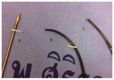
The high success and low complication rates of EUS-BD make it an appropriate technique for biliary access. Herein, we reported a special technique of using a self-made, tapered-tip, Teflon catheter for graded dilatation instead of using the cauterized instrument such as needle knife during enterobiliary tract creation to minimize the tissue damage and allow time for gradual expansion of the SEMS[10]. The cited study revealed high technical and clinical success rates of 100% and 91% respectively, with lower complications, only 6%, compared with the conventional methods by using a needle knife followed by balloon dilation with 85%-100% success rates and 5%-35% complication rates[2-8]. However, the self-made, tapered-tip, Teflon catheter for biliary access had limitations in some cases, and this instrument was not a commercial product; therefore, the use of alternative equipment should be considered. Interestingly, the Soehendra stent retriever has been used to retrieve the previous plastic stent while keeping the guidewire in place for easier stent exchange. Later on, some endoscopists reported the use of this instrument for negotiating the ERCP tract, such as biliary stent placement, in patients with very tight biliary and pancreatic duct strictures owing to benign or malignant obstruction[11]. Recently, some endosonographers also reported using it to create the neotract for EUS-guided drainage[12,13]. Regarding the characteristics of the tip of the Soehendra stent retriever - slim style (Figure 6) and ability to function as a drill, the authors tried using this instrument for neotract creation. In the present study, we demonstrated the feasibility of using the Soehendra stent retriever as a new instrument for neotract creation with the technical and clinical success rates of 100% and 91.7%, respectively, comparable to those of previous reports. In this case series, the authors used the Soehendra stent retriever for initial neotract creation by drilling mechanism and followed by Soehendra dilators for neotract dilatation. However, considering the mechanism of this instrument via the drilling effect, the authors realized that there could be more tissue damage compared to the simple graded dilatation effect. Although the complication reported in this series was higher(major 16% and minor 33%) than the previous one (which was only 6%), especially pneumoperitoneum, the authors showed that pneumoperitoneum, comprising one-third of the minor complications, spontaneously disappeared within a week and required no other interventions. Major complications occurred in 2 patients, including patient No. 4, who developed clinical signs of sepsis and localized peritonitis about 5 d after the procedure. A computed tomography scan of this patient showed accumulation of fluid next to the stomach; this fluid was successfully drained via a percutaneous technique, and it cultured positive for Enterococcus faecalis. The patient gradually recovered, and he was discharged 29 d after the procedure. Patient No. 10 developed upper gastrointestinal bleeding after the procedure; the bleeding was traced to a hepatic artery aneurysm, and it was successfully treated via angioembolisation[14]. Nevertheless, compared to other reports of the complications caused by EUS-BD, which accounted for 6%-35%[1-8], the complication rate using this instrument was still acceptable. Therefore, we recommend using the Soehendra stent retriever as a new alternative instrument for the creation and dilation of enterobiliary tract access in EUS-BD.
The authors endorse this instrument, which is a commercial product that might be benefit and adapted, as an alternative option for neotract creation with high technical and clinical success rates as well as a complication rate that did not differ from those of previous reports of EUS-BD, especially after failure to use a tapered-tip dilator prior to balloon dilation.
Endoscopic ultrasound-guided biliary drainage (EUS-BD) is considered as an alternative treatment for patients with difficult malignant biliary obstruction in whom endoscopic retrograde cholangiopancreatography (ERCP) for failed palliative biliary drainage. EUS-BD is currently comparative or superior to conventional percutaneous transhepatic biliary drainage in some retrospective studies. This procedure has been reported effective and safe when performed by experts. The important issues of high success and low complication rate of EUS-BD makes it an appropriate technique for biliary access. The techniques for biliary access were classified into neotract creation with cauterized or non-cauterized and neotract dilatation with graded or balloon dilation. Many studies have reported techniques and equipments used in these procedure. The authors usually do not use cauterization and balloon dilation for biliary access in EUS-BD due to concerns about complications, especially perforation and bile leakage. Despite experience with a non-cauterized and graded dilatation technique using the self-made, tapered-tip, Teflon catheter, which provided high success and low complication rates, there are still some limitations of these instruments in some particular cases.
Regarding the characteristics of the tip of Soehendra stent retriever which was slim including an ability to function as a drill. The authors demonstrated this particular instrument as an alternative option for neotract creation (non cauterization technique).
The Soehendra stent retriever was first used for biliary stent retrieval. It was recently proven useful in many therapeutic applications. The authors endorse this instrument, which is a commercial product that might be benefit and adapted, as an alternative option for neotract creation with high technical and clinical success rates of 100% and 91.7% respectively as well as a complication rate that did not differ from those of previous reports of EUS-BD.
Soehendra stent retriever is an alternative instrument for non-cauterized neotract creation technique in EUS-BD.
EUS-BD which are EUS guided-choledochoduodenostomy or EUS guided-hepaticogastrostomy, is considered an alternative treatment for patients with biliary obstruction in whom ERCP for failed palliative biliary drainage. The important systematic procedures were localization and puncture, followed by guidewire negotiation into the desired position, followed by biloenteric fistula creation and dilatation by using an over-the-guidewire technique, and finally stent placement.
Authors developed this non-cauterized and graded dilatation technique for biliary access in concern of complication, perforation and bile leakage, instead of knife and balloon dilatation method to create neotract. However the result of this study showed no advantage compared to old technique. However the use of commercial product may be a benefit and be adapted for other expert interventional endoscopist.
P- Reviewer: Cho HJ, Rabago L S- Editor: Yu J L- Editor: A E- Editor: Ma S
| 1. | Attasaranya S, Netinasunton N, Jongboonyanuparp T, Sottisuporn J, Witeerungrot T, Pirathvisuth T, Ovartlarnporn B. The Spectrum of Endoscopic Ultrasound Intervention in Biliary Diseases: A Single Center’s Experience in 31 Cases. Gastroenterol Res Pract. 2012;2012:680753. [RCA] [PubMed] [DOI] [Full Text] [Full Text (PDF)] [Cited by in Crossref: 34] [Cited by in RCA: 45] [Article Influence: 3.5] [Reference Citation Analysis (0)] |
| 2. | Levy MJ. Therapeutic endoscopic ultrasound for biliary and pancreatic disorders. Curr Gastroenterol Rep. 2010;12:141-149. [RCA] [PubMed] [DOI] [Full Text] [Cited by in Crossref: 4] [Cited by in RCA: 7] [Article Influence: 0.5] [Reference Citation Analysis (0)] |
| 3. | Kim TH, Kim SH, Oh HJ, Sohn YW, Lee SO. Endoscopic ultrasound-guided biliary drainage with placement of a fully covered metal stent for malignant biliary obstruction. World J Gastroenterol. 2012;18:2526-2532. [RCA] [PubMed] [DOI] [Full Text] [Full Text (PDF)] [Cited by in CrossRef: 44] [Cited by in RCA: 48] [Article Influence: 3.7] [Reference Citation Analysis (0)] |
| 4. | Itoi T, Sofuni A, Itokawa F, Tsuchiya T, Kurihara T, Ishii K, Tsuji S, Ikeuchi N, Umeda J, Moriyasu F. Endoscopic ultrasonography-guided biliary drainage. J Hepatobiliary Pancreat Sci. 2010;17:611-616. [RCA] [PubMed] [DOI] [Full Text] [Cited by in RCA: 2] [Reference Citation Analysis (0)] |
| 5. | Giovannini M, Bories E. EUS-Guided Biliary Drainage. Gastroenterol Res Pract. 2012;2012:348719. [RCA] [PubMed] [DOI] [Full Text] [Full Text (PDF)] [Cited by in Crossref: 31] [Cited by in RCA: 37] [Article Influence: 2.6] [Reference Citation Analysis (0)] |
| 6. | Irisawa A, Hikichi T, Shibukawa G, Takagi T, Wakatsuki T, Takahashi Y, Imamura H, Sato A, Sato M, Ikeda T. Pancreatobiliary drainage using the EUS-FNA technique: EUS-BD and EUS-PD. J Hepatobiliary Pancreat Surg. 2009;16:598-604. [RCA] [PubMed] [DOI] [Full Text] [Cited by in Crossref: 11] [Cited by in RCA: 16] [Article Influence: 1.0] [Reference Citation Analysis (0)] |
| 7. | Park do H, Jang JW, Lee SS, Seo DW, Lee SK, Kim MH. EUS-guided biliary drainage with transluminal stenting after failed ERCP: predictors of adverse events and long-term results. Gastrointest Endosc. 2011;74:1276-1284. [RCA] [PubMed] [DOI] [Full Text] [Cited by in Crossref: 220] [Cited by in RCA: 243] [Article Influence: 17.4] [Reference Citation Analysis (0)] |
| 8. | Tarantino I, Barresi L, Fabbri C, Traina M. Endoscopic ultrasound guided biliary drainage. World J Gastrointest Endosc. 2012;4:306-311. [RCA] [PubMed] [DOI] [Full Text] [Full Text (PDF)] [Cited by in CrossRef: 16] [Cited by in RCA: 19] [Article Influence: 1.5] [Reference Citation Analysis (0)] |
| 9. | Prachayakul V, Aswakul P, Kachinthorn U. Tapered-tip Teflon catheter: another device for sequential dilation in endoscopic ultrasound-guided hepaticogastrostomy. Endoscopy. 2011;43 Suppl 2 UCTN:E213-E214. [RCA] [PubMed] [DOI] [Full Text] [Cited by in Crossref: 6] [Cited by in RCA: 7] [Article Influence: 0.5] [Reference Citation Analysis (0)] |
| 10. | Prachayakul V, Aswakul P. A novel technique for endoscopic ultrasound-guided biliary drainage. World J Gastroenterol. 2013;19:4758-4763. [RCA] [PubMed] [DOI] [Full Text] [Full Text (PDF)] [Cited by in CrossRef: 24] [Cited by in RCA: 28] [Article Influence: 2.3] [Reference Citation Analysis (0)] |
| 11. | Kato H, Kawamoto H, Noma Y, Sonoyama T, Tsutsumi K, Fujii M, Okada H, Yamamoto K. Dilatation by Soehendra stent retriever is feasible and effective in multiple deployment of metallic stents to malignant hilar biliary strictures. Hepatogastroenterology. 2013;60:286-290. [PubMed] |
| 12. | Vila JJ, Goñi S, Arrazubi V, Bolado F, Ostiz M, Javier Jiménez F. Endoscopic ultrasonography-guided transgastric biliary drainage aided by Soehendra stent retriever. Am J Gastroenterol. 2010;105:959-960. [RCA] [PubMed] [DOI] [Full Text] [Cited by in Crossref: 7] [Cited by in RCA: 8] [Article Influence: 0.5] [Reference Citation Analysis (0)] |
| 13. | Yamaguchi T, Ishihara T, Tadenuma H, Kobayashi A, Nakamura K, Kouzu T, Saisho H. Use of a Soehendra stent retriever to treat a pancreatic pseudocyst with EUS-guided cystogastrostomy. Endoscopy. 2004;36:755. [PubMed] |
| 14. | Prachayakul V, Thamtorawat S, Siripipattanamongkol C, Thanathanee P. Bleeding left hepatic artery pseudoaneurysm: a complication of endoscopic ultrasound-guided hepaticogastrostomy. Endoscopy. 2013;45 Suppl 2 UCTN:E223-E224. [RCA] [PubMed] [DOI] [Full Text] [Cited by in Crossref: 7] [Cited by in RCA: 11] [Article Influence: 1.0] [Reference Citation Analysis (0)] |


