Copyright
©The Author(s) 2015.
World J Gastroenterol. Nov 21, 2015; 21(43): 12249-12260
Published online Nov 21, 2015. doi: 10.3748/wjg.v21.i43.12249
Published online Nov 21, 2015. doi: 10.3748/wjg.v21.i43.12249
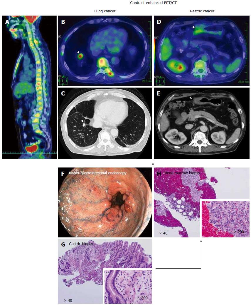
Figure 5 A case of synchronous disseminated carcinomatosis of the bone marrow associated with gastric cancer in a 60-year-old man: Findings of positron emission tomography/computed tomography imaging, endoscopic examination, and bone marrow biopsy.
History: The patient visited a local doctor in August 2007 with complaints of left chest and back pain. CT and magnetic resonance imaging revealed a “lung tumor” and “bone metastasis (thoracic vertebrae)”; therefore, the patient was referred to the Shikoku Cancer Center. Laboratory findings: Marked elevation of serum ALP (2594 U/L), CRP (34.74 mg/dL), and mild elevation of serum LDH (342 U/L) were observed with hematological disorder (DIC and elevated WBC [possible leukoerythroblastosis]). Tumor marker (CA19-9 162 U/mL) and bone metabolic markers (1CTP 8.9 ng/mL; BAP 258 μg/L) were elevated. These findings were suggestive of recurrence of gastric cancer, perhaps disseminated carcinomatosis of the bone marrow. PET/CT imaging: FDG accumulation was observed in all spinal vertebrae and the sternum in the sagittal view of the PET/CT fusion image (A). In the transaxial views of CT and PET/CT fusion images, a tumor mass with FDG accumulation was observed in the right lung (arrowheads; B, C) and thickening of the wall with FDG accumulation was observed in the gastric antrum (arrowheads; D, E). These findings on PET/CT suggested the presence of lung cancer together with gastric cancer. Endoscopic examination: Multiple erosions were observed in the gastric antrum/pyloric ring (F). In the tissue specimen obtained from the erosions, proliferation of signet-ring cell carcinoma was observed (G). Bone marrow biopsy: In order to determine the origin of the bone lesions, bone marrow biopsy was performed. Signet-ring cell carcinoma characterized by clear abundant cytoplasm and eccentrically positioned nuclei was found in the hematopoietic bone marrow (H). This histologic finding suggests the bone lesions originated from the gastric cancer. PET: Positron emission tomography; CT: Computed tomography.
- Citation: Iguchi H. Recent aspects for disseminated carcinomatosis of the bone marrow associated with gastric cancer: What has been done for the past, and what will be needed in future? World J Gastroenterol 2015; 21(43): 12249-12260
- URL: https://www.wjgnet.com/1007-9327/full/v21/i43/12249.htm
- DOI: https://dx.doi.org/10.3748/wjg.v21.i43.12249


