Copyright
©The Author(s) 2015.
World J Gastroenterol. Nov 7, 2015; 21(41): 11832-11841
Published online Nov 7, 2015. doi: 10.3748/wjg.v21.i41.11832
Published online Nov 7, 2015. doi: 10.3748/wjg.v21.i41.11832
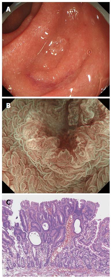
Figure 7 False-positive magnifying endoscopy with narrow-band imaging diagnosis.
A: Endoscopic findings using conventional endoscopy with white light imaging. A whitish, slightly depressed lesion (5 mm in diameter) is observed in the second portion of the duodenum. In this case, magnifying endoscopy with narrow-band imaging diagnosis (M-NBI) examination was conducted before biopsy; B: Endoscopic findings using M-NBI. A clear demarcation line (DL) is visible because of differences in the vessel plus surface (VS) component between the tumor and surrounding mucosa. V: The individual vessels show a variety of morphologies, such as open- and closed-looped and coil-shaped, with no two microvessels sharing the same morphology. The microvessels are anastomosing with each other within the intervening parts but show no consistent regularity. Therefore, this lesion was assessed as an irregular microvascular (MV) pattern; S: This individual section of marginal crypt epithelium (MCE) shows a curved morphology but lacks continuity or a consistent directionality, and the intervening parts are also irregular with unequal sizes. Therefore, this lesion was assessed as an irregular mucosal microsurface (MS) pattern. The VS classification of this lesion was an irregular MV pattern and irregular MS pattern with a DL. Therefore, the M-NBI diagnosis was cancer; C: The final histological diagnosis was a low-grade adenoma.
- Citation: Tsuji S, Doyama H, Tsuji K, Tsuyama S, Tominaga K, Yoshida N, Takemura K, Yamada S, Niwa H, Katayanagi K, Kurumaya H, Okada T. Preoperative endoscopic diagnosis of superficial non-ampullary duodenal epithelial tumors, including magnifying endoscopy. World J Gastroenterol 2015; 21(41): 11832-11841
- URL: https://www.wjgnet.com/1007-9327/full/v21/i41/11832.htm
- DOI: https://dx.doi.org/10.3748/wjg.v21.i41.11832


