Copyright
©The Author(s) 2015.
World J Gastroenterol. Nov 7, 2015; 21(41): 11832-11841
Published online Nov 7, 2015. doi: 10.3748/wjg.v21.i41.11832
Published online Nov 7, 2015. doi: 10.3748/wjg.v21.i41.11832
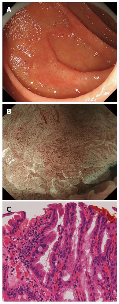
Figure 6 Duodenal adenocarcinoma with typical magnifying endoscopy with narrow-band imaging findings.
A: Endoscopic findings using conventional endoscopy with white light imaging. A reddish, slightly elevated lesion (13 mm in diameter, arrows) is observed in the second portion of the duodenum; B: Endoscopic findings using magnifying endoscopy with narrow-band imaging findings (M-NBI). A clear demarcation line (DL) is visible because of differences in the vessel plus surface (VS) component between the cancerous and noncancerous mucosa. V: Proliferation of microvessels with variable sizes, asymmetrical distribution and irregular arrangement make this an irregular microvascular (MV) pattern; S: There are areas where the marginal crypt epithelium (MCE) cannot be visualized and where the visible MCE shows a variety of morphologies, an asymmetrical distribution and an irregular arrangement. This lesion is assessed as an irregular mucosal microsurface (MS) pattern. The VS classification of this lesion was an irregular MV pattern and irregular MS pattern with a DL. Therefore, the M-NBI diagnosis was cancer; C: The final histological diagnosis was a well-differentiated intramucosal adenocarcinoma.
- Citation: Tsuji S, Doyama H, Tsuji K, Tsuyama S, Tominaga K, Yoshida N, Takemura K, Yamada S, Niwa H, Katayanagi K, Kurumaya H, Okada T. Preoperative endoscopic diagnosis of superficial non-ampullary duodenal epithelial tumors, including magnifying endoscopy. World J Gastroenterol 2015; 21(41): 11832-11841
- URL: https://www.wjgnet.com/1007-9327/full/v21/i41/11832.htm
- DOI: https://dx.doi.org/10.3748/wjg.v21.i41.11832


