Published online Sep 14, 2015. doi: 10.3748/wjg.v21.i34.9993
Peer-review started: March 27, 2015
First decision: May 18, 2015
Revised: June 2, 2015
Accepted: June 26, 2015
Article in press: June 26, 2015
Published online: September 14, 2015
Processing time: 173 Days and 16.5 Hours
AIM: To evaluate the detectability of gallbladder stones by dual-energy spectral computed tomography (CT) imaging.
METHODS: Totally 217 patients with surgically confirmed gallbladder stones were retrospectively analyzed who underwent single-source dual-energy CT scanning from August 2011 to December 2013. Polychromatic images were acquired. And post-processing software was used to reconstruct monochromatic (40 keV and 140 keV) images, and calcium-lipid pair-wise base substance was selected to acquire calcium base images and lipid base images. The above 5 groups of images were evaluated by two radiologists separately with 10-year experience in CT image reading. In the 5 groups of images, the cases in the positive group and negative group were counted and then the detection rate was calculated. The inter-observer agreement on the scoring results was analyzed by Kappa test, and the scoring results were analyzed by Wilcoxon test, with P < 0.05 indicating that the difference was statistically significant. The stone detection results of the 5 groups of images were analyzed by χ2 test.
RESULTS: There was good inter-observer agreement (κ= 0.772). In 217 patients with gallbladder stones, there was a statistically significant difference in stone visualization between spectral images (40 keV, 140 keV, calcium base and lipid base images) and polychromatic images (P < 0.05). 40 keV monochromatic images were better than 140 keV monochromatic images (4.90 ± 0.35 vs 4.53 ± 1.15, P < 0.05), and calcium base images were superior to lipid base images (4.91 ± 0.43 vs 4.77 ± 0.63, P < 0.05), but there was no statistically significant difference between 40 keV monochromatic images and calcium base images (4.90 ± 0.35 vs 4.91 ± 0.43, P > 0.05). In 217 gallbladder stone patients, there were 21, 3, 28, 5 and 12 negative stone cases in polychromatic images, 40 keV images, 140 keV images, calcium base images and lipid base images, respectively, and the differences among the five groups were statistically significant (P < 0.05).
CONCLUSION: Monochromatic images and base substance images have a good clinical prospect in the iso-density stone detection.
Core tip: In traditional computed tomography (CT), gallbladder stones are indicated directly by high-density or low-density stones and indirectly by dilation of the intrahepatic biliary duct, left and right hepatic ducts, biliary duct and gallbladder. However, it is difficult to diagnose iso-density gallbladder stones, because gallbladder stones and bile are difficultly distinguished by traditional CT due to a same density. The development of spectral imaging recoups the defects of traditional CT in the diagnosis of iso-density stones. In spectral imaging, base substance images and low keV images are both promising to improve the detection rate of gallbladder stones; they provide a new approach for the differential diagnosis of gallbladder stones, and the reliable information for the clinical treatment of gallbladder stones.
- Citation: Chen AL, Liu AL, Wang S, Liu JH, Ju Y, Sun MY, Liu YJ. Detection of gallbladder stones by dual-energy spectral computed tomography imaging. World J Gastroenterol 2015; 21(34): 9993-9998
- URL: https://www.wjgnet.com/1007-9327/full/v21/i34/9993.htm
- DOI: https://dx.doi.org/10.3748/wjg.v21.i34.9993
Gallbladder stone disease is a common disorder and often accompanied by biliary stenosis or variation, and it seriously threatens the public health and quality of life. With the improvement of people’s living level, the morbidity of gallbladder stones trends to increase continuously[1]. In traditional computed tomography (CT), gallbladder stones are indicated directly by high-density or low-density stones and indirectly by dilation of the intrahepatic biliary duct, left and right hepatic ducts, biliary duct and gallbladder. Based on density, gallbladder stones are classified into: (1) high-density stones; (2) iso-density stones; (3) low-density stones; and (4) mixed-density stones[2]. Gallbladder stones are variable in shape, size and number. Solitary gallbladder stones or multiple gallbladder stones may occur. Gallbladder stones may have different sizes and a non-uniform density. Solitary stones are generally big; partial stones have a low density in the center and a high density in the peripheral area, demonstrating concentric circle changes; sandy stones are often deposited at the bottom of gallbladder, have a high density, and form the fluid level with bile at the upper part, and their diagnosis can be made by stone shift in the scanning with a changeable position. In a part of patients, gallbladder is enlarged with a long diameter > 5 cm, the gallbladder wall is thickened to be > 0.3 cm, and there is a low-density ring surrounding the gallbladder, which is the manifestation of secondary inflammatory edema[2]. However, it is difficult to diagnose iso-density gallbladder stones, because gallbladder stones and bile are difficultly distinguished by traditional CT due to a same density, which is troublesome for clinical treatment[3]. The development of spectral imaging recoups the defects of traditional CT in the diagnosis of iso-density stones. In spectral imaging, base substance images and low keV images are both promising to improve the detection rate of gallbladder stones; they provide a new approach for the differential diagnosis of gallbladder stones, and the reliable information for the clinical treatment of gallbladder stones.
This retrospective study was approved by the Ethics Committee in our hospital. Totally 217 patients with surgically confirmed gallbladder stones were retrospectively analyzed who underwent HDCT scanning for suspected gallbladder stones according to clinical symptoms and ultrasonic reports at our hospital from August 2011 to December 2013. There were 97 males and 120 females, with a mean age of 62 ± 15 (10-96) years.
HD CT750 Discovery was used for plain scanning under spectral imaging mode. The patients was in ≥ 6 h fasting before scanning. CT plain scanning was performed routinely in a dorsal position. After acquisition of anterior-posterior positioning images, the upper abdomen plain scanning was performed with a range from the top of the liver to the middle poles of bilateral kidneys. The scanning parameters were as follows: instantaneous 40-140 kVp rapid switching; tube current: 550 mA; slice thickness and interval: 5 mm; rotation speed: 0.8 r/s; and pitch: 1.375.
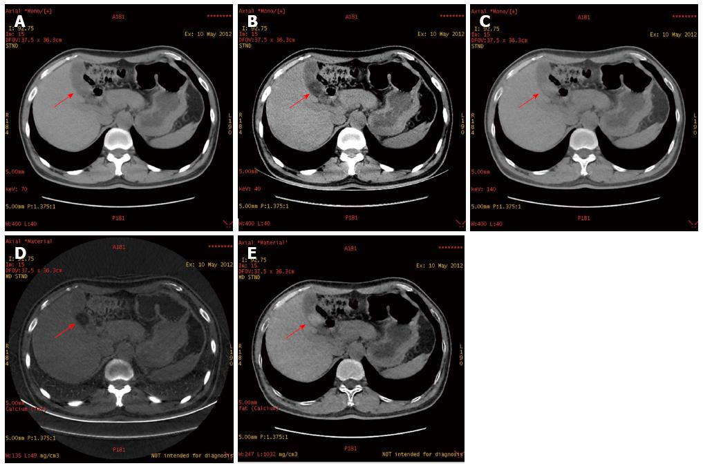
The image post-processing and evaluation were both performed with post-processing software on the professional workstation AW4.5 (GE HealthCare, United States) under a precondition of blinding to the final diagnosis results. Polychromatic images, 40 keV and 140 keV images were acquired. The calcium-lipid pair-wise base substance imaging was performed to acquire calcium base images and lipid base images. The gallbladder stone visualization in 5 groups of images was evaluated by two radiologists separately with 10-year experience in CT image reading. The following scorings were performed according to the density contrast resolution of gallbladder stones and bile: 1, no visualization; 2, poor visualization; 3, ordinary visualization; 4, good visualization; and 5, clear visualization. The images with a stone visualization score = 1-3 served as a negative group, and those with a stone visualization score = 4-5 served as a positive group. In 5 groups of images, the cases in the positive group and negative group were counted and then the detection rate was calculated.
SPSS 17.0 analysis software was used for statistical analyses: (1) the inter-group agreement on the scoring results was analyzed by Kappa test; (2) the scoring results were analyzed by Wilcoxon test, and P < 0.05 suggested a statistically significant difference; and (3) the detection results in 5 groups of images were analyzed by χ2 test, and P < 0.05 suggested a statistically significant difference.
The statistical analysis of observation results showed good inter-group agreement (κ= 0.772). In 217 gallbladder stone patients, there was a statistically significant difference in stone visualization between spectral images (40 keV, 140 keV, calcium base and lipid base images) and polychromatic images (P < 0.05) (Figure 1 and Table 1), and the 40 keV, calcium base and lipid base images were superior to the later, but the 140 keV imagings were inferior to polychromatic images.
| Image | Polychromatic | 40 keV | 140 keV | Calcium base | Lipid base |
| Mean | 4.62 | 4.90 | 4.53 | 4.91 | 4.77 |
| SD | 1.00 | 0.35 | 1.15 | 0.43 | 0.63 |
| P value | 0.00 | 0.00 | 0.00 | 0.00 |
Forty keV monochromatic images were better than 140 keV monochromatic images (4.90 ± 0.35 vs 4.53 ± 1.15, P < 0.05), and calcium base images were superior to lipid base images (4.91 ± 0.43 vs 4.77 ± 0.63, P < 0.05), but there was no statistically significant difference between 40 keV monochromatic images and calcium base images (4.90 ± 0.35 vs 4.91 ± 0.43, P > 0.05). Compared with polychromatic images, the stone visualization scores of 40 keV images and calcium base images were increased (Table 2).
| Polychromatic CT score | Spectral CT score | Number of 40 keV patients | Percentage of 40 keV patients | Number of calcium base patients | Percentage of calcium base patients |
| 1 | 2 | 0 | 0.00% | 1 | 0.46% |
| 1 | 3 | 1 | 0.46% | 1 | 0.46% |
| 1 | 4 | 4 | 1.84% | 2 | 0.92% |
| 1 | 5 | 4 | 1.84% | 5 | 2.30% |
| 2 | 3 | 2 | 0.92% | 1 | 0.46% |
| 2 | 4 | 4 | 1.84% | 1 | 0.46% |
| 2 | 5 | 2 | 0.92% | 4 | 1.84% |
| 3 | 4 | 3 | 1.38% | 1 | 0.46% |
| 3 | 5 | 1 | 0.46% | 3 | 1.38% |
| 4 | 5 | 9 | 4.15% | 11 | 5.07% |
| ≤ 3 | > 3 | 14 | 6.45% | 14 | 6.45% |
In 217 gallbladder stone patients, there were 21, 3, 28, 5 and 12 negative stone cases in polychromatic images, 40 keV images, 140 keV images, calcium base images and lipid base images, respectively (Table 3). The differences in the negative stone cases among the five groups were statistically significant (P < 0.05), and the detection rates of stones by 40 keV and calcium base images were higher.
| Group (score) | Polychromatic images | 40 keV images | 140 keV images | Calcium base images | Lipid base images |
| Negative group (1-2) | 21 (9.68) | 3 (1.38) | 28 (12.90) | 5 (2.30) | 12 (5.53) |
| Positive group (3-5) | 196 (90.32) | 214 (98.62) | 189 (87.10) | 212 (97.70) | 205 (94.47) |
| Total | 217 | 217 | 217 | 217 | 217 |
Cholestasis: Sitting meditation habit, obesity, pregnancy, biliary obstruction or Oddi’s sphincter dysfunciton can cause a decrease of gallbladder muscular tension and a delay of emptying, thus inducing cholestasis. When cholestasis occurs, the bile is detained in the gallbladder, and the water resorption is increased so that the bile is excessively concentrated. The chemical stimulation of bile salt component can induce mucosal inflammation and thus change the absorption function. When bile salts are detained, the bile basicity is increased, and the capacity of bile salts to dissolve cholesterol is thereby decreased. All the above factors can influence the normal proportions of bile components, which is beneficial for gallbladder stone formation.
Biliary infection: The biliary mucosa has inflammation due to the chemical stimulation of concentrated bile or refluxed pancreatic juice and thus becomes susceptible to secondary bacterial infection which will aggravate inflammation. The causes of inflammation contribution to gallbladder stone formation are below: (1) Bile salts can reduce the surface tension and they are potent bile solvents. When gallbladder mucosal inflammation happens, bile salts in the bile are decreased. In addition, inflammation and infection can inhibit hepatocyte function to reduce the secretion of bile salts, thus the normal proportions of bile salts and cholesterol in the bile are decreased and resultantly cholesterol is easily deposited to form stones; (2) When inflammation occurs, a lot of proteins and calcium exude, thus promoting the precipitation of calcium bilirubinate; and (3) When the concentration of cholesterol or calcium bilirubinate in the bile is increased and the bile flow is slow, the inflammatory exudates (e.g., precipitated mucus), or fibrins, white blood cells, exfoliated epithelial cells, and aggregated bacteria often form the core of stone.
Cholesterol proportion imbalance: Cholesterol is increased for some reasons while bile salts are decreased, so that cholesterol is precipitated and accumulated to form a stone. Therefore, the main causes of gallbladder stone formation are sitting meditation habit, obesity, pregnancy, biliary obstruction or biliary infection[4].
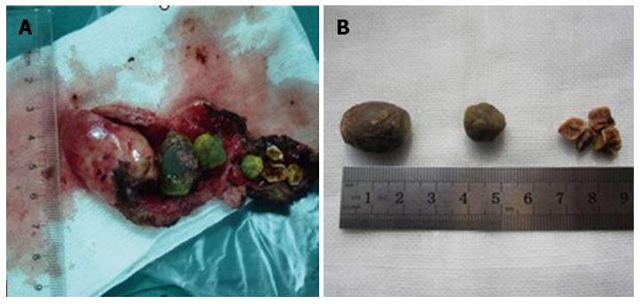
Gallbladder stones may be asymptomatic in the whole life, and it is mainly defined as a stone or stones found by ordinary examinations, also known as silent stones. Gallbladder stones also may cause biliary colic, and the presence of symptoms is associated with stone size and position, whether to result in obstruction, whether to have inflammation, and gallbladder function state (Figure 2). Symptomatic gallbladder stones primarily manifest as sudden, paroxysmal, severe right upper abdominal pain, with right upper abdominal discomfort during intermission, which may be relieved suddenly by a change of position. Abdominal pain is accompanied by nausea and vomiting, and often radiates to right shoulder and back, right chest and right sternum. According to the severity of stone obstruction and the severity and range of biliary infection, common bile duct stones may be asymptomatic or typical Charot’s triad (abdominal pain, shiver and ardent fever, and jaundice), or even acute obstructive suppurated cholangitis, or Charot’s pentalogy (abdominal pain, shiver & ardent fever, jaundice, shock, and mental disorder)[5].
In CT diagnosis, gallbladder stones are indicated directly by high-density or low-density stones and indirectly by dilation of the intrahepatic biliary duct, left and right hepatic ducts, biliary duct and gallbladder. Based on density, gallbladder stones are classified into: (1) high-density stones; (2) iso-density stones; and (3) low-density stones[2]. CT has a high resolution to characterize tissue density, and it can clearly show high-density or low-density biliary stones and distinguish cholesterol stones, bilirubin stones and mixed stones according to stone density[6]. CT value is a uniform unit reflecting tissue density, with water CT value set as 0 and bile CT value as 0-20 Hu. The stones with the same density as bile cannot be visualized, so traditional CT scanning cannot determine the presence of such stones; these stones through which X-ray can penetrate are called negative stones. Besides, individual stones can be visualized as filling defect shadows with a density slightly lower than bile in CT images and measured with a negative value 20 Hu lower than bile or < 0, which are known as low-density stones. With the bile density of 0-20 Hu as a threshold, those stones with a CT value > 20 Hu are high-density stones. Stone CT value is measured by calcium quantification analysis, and a high calcium content indicates a high density of stone and the image is clear; in contrast, a low calcium content indicates a low density of stone and the image is vague[7].
In traditional CT, X-ray generated by the X-ray tube has a continuous energy distribution, and X-ray for imaging is polychromatic so that the obtained traditional CT images reflect the average polychromatic effect. Influenced by such average effect, different substances have a similar CT value in many cases, which cannot be distinguished[8-10]. With the development of CT technology in recent years, the emergence of spectral images improves the detection rate of diseases and offsets some defects of traditional CT. In spectral CT imaging, energy resolution and physical and chemical property resolution are added on the basis of spatial resolution and temporal resolution. These relatively pure monochromatic images can greatly reduce the effects of hardening artifacts and provide the relatively pure CT value images. In other words, CT value is more consistent and reliable at different positions in the whole field, in different scans, or in different patients[8-11]. The attenuation of X-ray passing through a substance can objectively reflect the energy of X-ray. X-ray absorption coefficient of any substance can be determined by the absorption coefficients of two base substances, so the attenuation of one substance can be converted into the densities of two substances producing the same attenuation, achieving the separation of substance components and substances. This is a physical foundation for substance separation[12-15]. We observed 5 image groups of gallbladder stones (40 keV, polychromatic images, 140 keV, calcium base and lipid base images), and improved the resolution of images acquired from low-density or iso-density gallbladder stone patients whose diagnosis was easily missed by image debugging and using different base substance imaging. As a result, the detection rate of gallbladder stones was increased and the patients received the effective treatment.
In this study, we compared the scores of stone visualization of polychromatic CT images and spectral images acquired from 217 stone patients, and found that the scores of spectral images were higher than those of polychromatic CT images. It suggests that spectral imaging can visualize stones better. According to the comparison between the negative group and positive group of stones, the detection rate of negative stones was greater by spectral images than by polychromatic CT. In the present study, high-density stones and low-density stones could be well visualized in polychromatic CT images, so the stones in the positive group were mainly high-density stones and low-density stones, and the stones in the negative group were isodensity stones. Therefore, spectral imaging can improve the detection rate of negative stones (isodensity stones).
In conclusion, monochromatic images and base substance images are advantageous over polychromatic CT images in gallbladder stone visualization, especially negative stones (iso-density stones), which cannot be shown in polychromatic CT images. Therefore, base substance images and optimal low keV images have a good clinical prospect in the detection of iso-density stones. However, iso-density stones have a low incidence and only a few of cases are available, so future studies with a big sample size of iso-density stones are needed.
It is difficult to diagnose iso-density gallbladder stones, because gallbladder stones and bile are difficultly distinguished by traditional computed tomography (CT) due to a same density.
The development of spectral imaging recoups the defects of traditional CT in the diagnosis of iso-density stones.
In spectral imaging, base substance images and low keV images are both promising to improve the detection rate of gallbladder stones.
Authors provide a new approach for the differential diagnosis of gallbladder stones, and the reliable information for the clinical treatment of gallbladder stones.
Gallbladder stone disease is a common disorder and often accompanied by biliary stenosis or variation, and it seriously threatens the public health and quality of life. In traditional CT, gallbladder stones are indicated directly by high-density or low-density stones and indirectly by dilation of the intrahepatic biliary duct, left and right hepatic ducts, biliary duct and gallbladder.
This article is interesting. There are 1.3%-13% of false-negatives because X-ray do not contrast with pure cholesterol gallstones and also with black pigment gallstones and some brown pigment stones. Gallstone disease has afflicted humans since the time of Egyptian kings, and gallstones have been found during autopsies on mummies. Gallstone prevalence in adult population ranges from 10% to 15%. Ultrastructural analysis by scanning electron microscopy is useful in the classification and study of pigment gallstones. Moreover, X-ray diffractometry analysis and infrared spectroscopy of gallstones are of fundamental importance for an accurate stone analysis. An accurate study of gallstones is useful to understand gallstone pathogenesis.
P- Reviewer: Cariati A, Wang YF S- Editor: Yu J L- Editor: Wang TQ E- Editor: Ma S
| 1. | Cui Y, Li Z, Zhao E, Cui N. Risk factors in patients with hereditary gallstones in Chinese pedigrees. Med Princ Pract. 2012;21:467-471. [RCA] [PubMed] [DOI] [Full Text] [Cited by in Crossref: 1] [Cited by in RCA: 3] [Article Influence: 0.2] [Reference Citation Analysis (0)] |
| 2. | Xu SQ, Zhao LF. CT Diagnosis of Cholelithiasis. Shiyong Linchuang Yiyao Zazhi. 2009;13:128-130. |
| 3. | Bang BW, Hong JT, Choi YC, Jeong S, Lee DH, Kim HK, Park SG, Jeon YS. Is endoscopic ultrasound needed as an add-on test for gallstone diseases without choledocholithiasis on multidetector computed tomography? Dig Dis Sci. 2012;57:3246-3251. [RCA] [PubMed] [DOI] [Full Text] [Cited by in Crossref: 5] [Cited by in RCA: 4] [Article Influence: 0.3] [Reference Citation Analysis (0)] |
| 4. | Liu XJ. Causes and Prevention of Gallbladder Stone. Henan Yufang Yixue Zazhi. 2003;14:90. |
| 5. | Li GZ, Dai JP, Wang YS. Clinical CT Diagnostics. Beijing: China Science and Technology Press 1997; 448. |
| 6. | Anderson SW, Lucey BC, Varghese JC, Soto JA. Accuracy of MDCT in the diagnosis of choledocholithiasis. AJR Am J Roentgenol. 2006;187:174-180. [RCA] [PubMed] [DOI] [Full Text] [Cited by in Crossref: 56] [Cited by in RCA: 58] [Article Influence: 3.1] [Reference Citation Analysis (0)] |
| 7. | Zhang YL. Study on Relationship between CT Imaging Manifestations and Chemical Type of Gallbladder Stone. Zhongguo Linchuang Yixue Yingxiang. 1998;9:209-210. |
| 8. | Karçaaltıncaba M, Aktaş A. Dual-energy CT revisited with multidetector CT: review of principles and clinical applications. Diagn Interv Radiol. 2011;17:181-194. [RCA] [PubMed] [DOI] [Full Text] [Cited by in Crossref: 26] [Cited by in RCA: 75] [Article Influence: 5.0] [Reference Citation Analysis (0)] |
| 9. | Remy-Jardin M, Faivre JB, Pontana F, Molinari F, Tacelli N, Remy J. Thoracic applications of dual energy. Semin Respir Crit Care Med. 2014;35:64-73. [RCA] [PubMed] [DOI] [Full Text] [Cited by in Crossref: 26] [Cited by in RCA: 19] [Article Influence: 1.7] [Reference Citation Analysis (0)] |
| 10. | Kang MJ, Park CM, Lee CH, Goo JM, Lee HJ. Dual-energy CT: clinical applications in various pulmonary diseases. Radiographics. 2010;30:685-698. [RCA] [PubMed] [DOI] [Full Text] [Cited by in Crossref: 138] [Cited by in RCA: 142] [Article Influence: 9.5] [Reference Citation Analysis (0)] |
| 11. | Ren QG, Hua YQ, Li JY. The basic principle and clinical applications of CT spectral imaging. Guoji Yixue Fangshexue Zazhi. 2011;34:559-563. |
| 12. | Lin XZ, Shen Y, Chen KM. Basic Principle and Research Progress of Clinical Application of CT Spectral Imaging. Zhonghua Fangshexue Zazhi. 2011;45:798-800. |
| 13. | Lin XZ, Wu ZY, Li WX, Zhang J, Xu XQ, Chen KM, Yan FH. Differential diagnosis of pancreatic serous oligocystic adenoma and mucinous cystic neoplasm with spectral CT imaging: initial results. Clin Radiol. 2014;69:1004-1010. [RCA] [PubMed] [DOI] [Full Text] [Cited by in Crossref: 14] [Cited by in RCA: 18] [Article Influence: 1.6] [Reference Citation Analysis (0)] |
| 14. | Hao L, Liu AL, Wang HQ, Zhang T, Liu JH, Wang S. Spectral CT imaging in differential diagnosis of posterior bladder wall cancer and prostatic hyperplasia into bladder. Zhongguo Yixue Yingxiang Jishu. 2013;29:269-272. |
| 15. | Lv P, Lin XZ, Li J, Li W, Chen K. Differentiation of small hepatic hemangioma from small hepatocellular carcinoma: recently introduced spectral CT method. Radiology. 2011;259:720-729. [RCA] [PubMed] [DOI] [Full Text] [Cited by in Crossref: 152] [Cited by in RCA: 167] [Article Influence: 11.9] [Reference Citation Analysis (0)] |


