Published online Sep 14, 2015. doi: 10.3748/wjg.v21.i34.10041
Peer-review started: March 3, 2015
First decision: April 13, 2015
Revised: April 27, 2015
Accepted: June 10, 2015
Article in press: June 10, 2015
Published online: September 14, 2015
Processing time: 205 Days and 7.1 Hours
Here we present the case of a 64-year-old female with a duodenal carcinoid tumor treated by ligation-assisted endoscopic submucosal resection (ESMR-L) with circumferential mucosal incision (CMI). Band ligation was effective in resecting the duodenal carcinoid tumor after CMI, with an uneventful post-procedural course. Histopathological examination showed clear tumor margins at deeper tissue levels. Thus, in the present case, ESMR-L with CMI was useful for the treatment of duodenal carcinoid tumor.
Core tip: This report presents the case of a 64-year-old female with duodenal carcinoid tumor treated by ligation-assisted endoscopic submucosal resection (ESMR-L) with circumferential mucosal incision (CMI). Band ligation was effective in resecting the duodenal carcinoid tumor after CMI. The post-procedural course was uneventful without bleeding and perforation. Histopathological examination showed clear tumor margins at deeper tissue levels. Thus, ESMR-L with CMI can be effective for the management of duodenal carcinoid tumors.
- Citation: Harada H, Suehiro S, Shimizu T, Katsuyama Y, Hayasaka K, Ito H. Ligation-assisted endoscopic submucosal resection with circumferential mucosal incision for duodenal carcinoid tumor. World J Gastroenterol 2015; 21(34): 10041-10044
- URL: https://www.wjgnet.com/1007-9327/full/v21/i34/10041.htm
- DOI: https://dx.doi.org/10.3748/wjg.v21.i34.10041
Carcinoid tumors are relatively rare, slow-growing tumors that show low-grade atypia. Typically, gastrointestinal carcinoid tumors are found incidentally during screening esophagogastroduodenoscopy (EGD) and colonoscopy[1], with the most common location being the rectum, followed by the stomach, duodenum, and appendix. Carcinoid tumors also have excellent 5-year survival rates of 98.9%-100% after curative resection, provided there is no vessel infiltration, they are within the submucosa, and they measure < 10 mm in diameter[2-5]. Therefore, conventional endoscopic mucosal resection (EMR) is usually sufficient for the resection of small submucosal carcinoid tumors diagnosed by endoscopic ultrasound (EUS). However, those at deeper locations within the submucosa are not always amenable to complete resection by conventional EMR.
Some studies have recently reported that ligation-assisted endoscopic submucosal resection (ESMR-L) and endoscopic submucosal dissection (ESD) can facilitate complete resection of carcinoid tumors[6-8]. Furthermore, the efficacy of ligation-assisted endoscopic enucleation (EE-L) has been reported for the treatment of submucosal tumors (e.g., leiomyomas and gastrointestinal stromal tumors)[9]. However, in cases where the submucosa has significant fibrosis, it may be impossible to dissect the fibrosis by ESD alone or to suck the tumor through a ligation device. In this setting, ESMR-L with circumferential mucosal incision (CMI) may be effective. In this report, we therefore describe our positive experience using this technique to treat a duodenal carcinoid tumor identified by mass screening EGD.
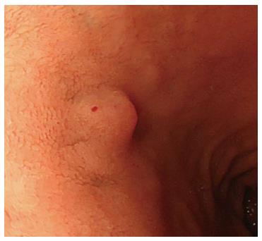
A 64-year-old female underwent a screening EGD that revealed a 7-mm submucosal tumor in the anterior wall of the duodenal bulb (Figure 1). EUS revealed the lesion to be a 6 mm × 5 mm hypoechoic, uniform tumor with a regular margin within the third layer. Therefore, we performed ligation-assisted ESMR with CMI after obtaining written informed consent from the patient.
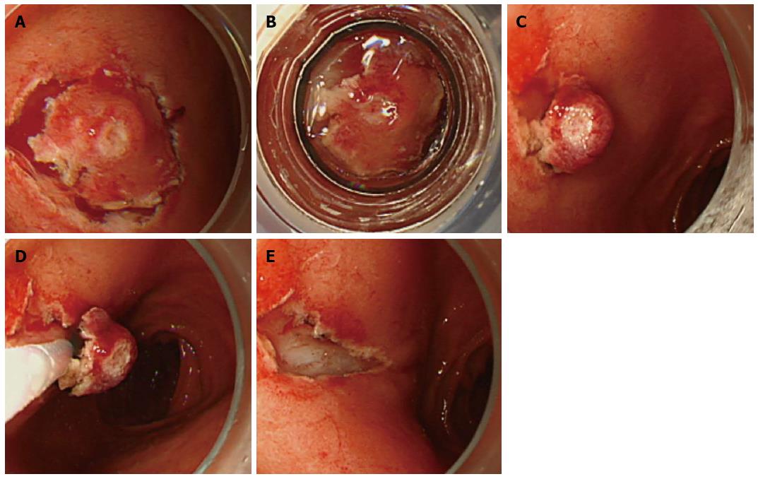
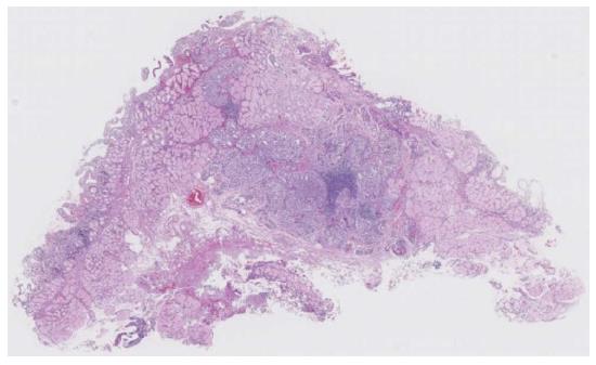
The procedure was performed as follows. Firstly, to elevate the tumor, we injected sodium hyaluronate in the underlying submucosa. After lifting the mucosa, we used a FlushKnife BT (DK2618JB; Fujifilm, Tokyo, Japan) to make a hemicircumferential mucosal incision around the tumor. Although we attempted ESD, it was not possible because of fibrosis. Therefore, in addition to the hemicircumferential mucosal incision (Figure 2A), we suctioned the tumor with a ligation device (pneumo-activate EVL device; Sumitomo Bakelite, Tokyo, Japan) (Figure 2B), which changed the shape of the tumor to a pseudopolyp that was suitable for band ligation (Figure 2C). Next, we performed polypectomy beneath the rubber band using a snare in ENDOCUT mode (Figures 2D and E). Histopathological examination revealed a G1 neuroendocrine tumor measuring 5.5 mm × 4.6 mm with clear tumor margins at deep tissue levels (the tumor cells were positive for chromogranin-A, synaptophysin, and CD56) (Figure 3). The post-procedural course was uneventful without bleeding and perforation.
Recently, the efficacy of ESD has been reported for carcinoid tumors that have not spread beyond the submucosa. However, as observed in the present case, submucosal dissection may be impossible when complicated by severe fibrosis. Although histopathological examination of the resected tumor failed to show severe fibrosis, we believe this resulted from the influence of hyperplasia of Brunner’s glands. It is believed that submucosal injection is particularly difficult in the duodenal bulb in comparison with application in other parts of the duodenum because of the presence of many Brunner’s glands.
Another important consideration is the report by Shida et al[10] who performed ESD with a ligation device (ESD-L). In our opinion, naming the procedure as ESD-L is not appropriate because it is not performed to dissect the submucosa. Consequently, we would argue that ESMR-L with CMI is more appropriate.
Band ligation was effective for resecting the duodenal carcinoid tumor after CMI because it was relatively easy to center and suction the duodenal carcinoid tumor with band ligation. However, band ligation can cause mucosal bleeding, making it difficult to ensure appropriate suction at the center of the tumor without CMI. Therefore, it is possible that band ligation after CMI facilitates easy suction of the submucosal tumor.
In ESMR-L with CMI, the tumor is cut by snaring beneath the rubber band after band ligation. Therefore, perforation is possible if the muscularis is involved, making it necessary to identify such involvement. This is particularly likely when band ligation involves the muscularis of the thin duodenal wall. Thus, if the ligation band involves the muscularis, the lesion should be cut over the rubber band to avoid perforation, although this may affect histopathological examination.
The advantage of ESD is that tumors can be resected endoscopically, en bloc, and regardless of their size or the presence of fibrosis. However, considerable skill is needed to maintain an appropriate line of dissection when there is extensive fibrosis of the submucosa. In such a setting, there is an increased risk of exposure of the lower tumor margin at the time of dissection, which can influence the histopathological evaluation. In contrast, ESMR-L with CMI ensures that tumor involvement in the deeper tissue level is included.
In this case, ESMR-L with CMI had minimal burning effects on the cutting margin because the tumor was cut by snare beneath the rubber band. However, during ESD, the dissection line is precisely determined under direct vision and may prove to be superior to ESMR-L with CMI for curative resection and histopathological examination. Therefore, further investigation is warranted to compare these procedures.
We would like to thank Dr. Okamoto S, Department of Pathology, New Tokyo Hospital, for his pathological advice.
A 64-year-old female with a submucosal tumor in the duodenal bulb on the anterior wall.
A submucosal tumor was incidentally found by screening esophagogastroduodenoscopy.
Gastrointestinal stromal tumor, liposarcoma, angiosarcoma, granular cell tumor, and Brunner’s glands.
WBC 6.8 k/μL; HGB 13.50 g/dL; Gastrin 115 pg/mL; no remarkable findings for the other laboratory tests.
Esophagogastroduodenoscopy revealed a submucosal tumor in the duodenal bulb on the anterior wall. Endoscopic ultrasound revealed a hypoechoic and uniform tumor, 6 mm × 5 mm diameter, with a regular margin within the third layer.
Histopathological examination showed carcinoid of duodenum, positive for chromogranin-A, synaptophysin, and CD56.
The patient was treated by ligation-assisted endoscopic submucosal resection with circumferential mucosal incision.
Few cases of ligation-assisted endoscopic submucosal resection with circumferential mucosal incision for carcinoid tumor have been reported in the literature.
Carcinoid tumor, also called neuroendocrine tumor, is a relatively rare tumor which is occasionally found during screening endoscopy.
This case report described ligation-assisted endoscopic submucosal resection with circumferential mucosal incision for duodenal carcinoid. Band ligation was effective in resecting the duodenal carcinoid tumor after circumferential mucosal incision.
The authors have described a case of endoscopic resection for duodenal carcinoid tumor. The article highlights that band ligation facilitated complete resection with sucking the tumor after circumferential mucosal incision.
P- Reviewer: Kirsaclioglu CT, Niimi K S- Editor: Yu J L- Editor: Logan S E- Editor: Liu XM
| 1. | Caldarola VT, Jackman RJ, Moertel CG, Dockerty MB. Carcinoid tumors of the rectum. Am J Surg. 1964;107:844-849. [RCA] [PubMed] [DOI] [Full Text] [Cited by in Crossref: 76] [Cited by in RCA: 63] [Article Influence: 1.0] [Reference Citation Analysis (0)] |
| 2. | Soga J. Early-stage carcinoids of the gastrointestinal tract: an analysis of 1914 reported cases. Cancer. 2005;103:1587-1595. [RCA] [PubMed] [DOI] [Full Text] [Cited by in Crossref: 234] [Cited by in RCA: 220] [Article Influence: 11.0] [Reference Citation Analysis (0)] |
| 3. | Konishi T, Watanabe T, Kishimoto J, Kotake K, Muto T, Nagawa H. Prognosis and risk factors of metastasis in colorectal carcinoids: results of a nationwide registry over 15 years. Gut. 2007;56:863-868. [RCA] [PubMed] [DOI] [Full Text] [Cited by in Crossref: 179] [Cited by in RCA: 179] [Article Influence: 9.9] [Reference Citation Analysis (0)] |
| 4. | Modlin IM, Oberg K, Chung DC, Jensen RT, de Herder WW, Thakker RV, Caplin M, Delle Fave G, Kaltsas GA, Krenning EP. Gastroenteropancreatic neuroendocrine tumours. Lancet Oncol. 2008;9:61-72. [RCA] [PubMed] [DOI] [Full Text] [Cited by in Crossref: 1268] [Cited by in RCA: 1183] [Article Influence: 69.6] [Reference Citation Analysis (0)] |
| 5. | Onozato Y, Kakizaki S, Iizuka H, Sohara N, Mori M, Itoh H. Endoscopic treatment of rectal carcinoid tumors. Dis Colon Rectum. 2010;53:169-176. [RCA] [PubMed] [DOI] [Full Text] [Cited by in Crossref: 71] [Cited by in RCA: 72] [Article Influence: 4.8] [Reference Citation Analysis (0)] |
| 6. | Ono A, Fujii T, Saito Y, Matsuda T, Lee DT, Gotoda T, Saito D. Endoscopic submucosal resection of rectal carcinoid tumors with a ligation device. Gastrointest Endosc. 2003;57:583-587. [RCA] [PubMed] [DOI] [Full Text] [Cited by in Crossref: 120] [Cited by in RCA: 107] [Article Influence: 4.9] [Reference Citation Analysis (0)] |
| 7. | Lee DS, Jeon SW, Park SY, Jung MK, Cho CM, Tak WY, Kweon YO, Kim SK. The feasibility of endoscopic submucosal dissection for rectal carcinoid tumors: comparison with endoscopic mucosal resection. Endoscopy. 2010;42:647-651. [RCA] [PubMed] [DOI] [Full Text] [Cited by in Crossref: 86] [Cited by in RCA: 104] [Article Influence: 6.9] [Reference Citation Analysis (0)] |
| 8. | Yamaguchi N, Isomoto H, Nishiyama H, Fukuda E, Ishii H, Nakamura T, Ohnita K, Hayashi T, Kohno S, Nakao K. Endoscopic submucosal dissection for rectal carcinoid tumors. Surg Endosc. 2010;24:504-508. [RCA] [PubMed] [DOI] [Full Text] [Cited by in Crossref: 37] [Cited by in RCA: 37] [Article Influence: 2.3] [Reference Citation Analysis (0)] |
| 9. | Guo J, Liu Z, Sun S, Wang S, Ge N, Liu X, Wang G, Yang X. Ligation-assisted endoscopic enucleation for the diagnosis and resection of small gastrointestinal tumors originating from the muscularis propria: a preliminary study. BMC Gastroenterol. 2013;13:88. [RCA] [PubMed] [DOI] [Full Text] [Full Text (PDF)] [Cited by in Crossref: 8] [Cited by in RCA: 12] [Article Influence: 1.0] [Reference Citation Analysis (0)] |
| 10. | Shida T, Aminaka M, Shirai Y, Okimoto K, Tsuruta S, Kita E, Tsuchiya S, Kato K, Takahashi M. Endoscopic submucosal dissection with a ligation device for the treatment of rectal carcinoid tumor. Endoscopy. 2012;44 Suppl 2 UCTN:E4-E5. [RCA] [PubMed] [DOI] [Full Text] [Cited by in Crossref: 2] [Cited by in RCA: 2] [Article Influence: 0.2] [Reference Citation Analysis (0)] |


