Copyright
©The Author(s) 2015.
World J Gastroenterol. Apr 28, 2015; 21(16): 5099-5104
Published online Apr 28, 2015. doi: 10.3748/wjg.v21.i16.5099
Published online Apr 28, 2015. doi: 10.3748/wjg.v21.i16.5099
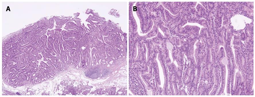
Figure 4 Pathologic examinations.
A: Hematoxylin and eosin staining showed proliferation of glands with complex architecture, situated deep in the mucosal layer with surface involvement (× 40); B: A higher magnification showed complex growth of glands lined with one to two layers of oxyntic epithelium with abundant chief cells. The lining cells showed moderate nuclear pleomorphism, but no mitotic figures were identified (× 200).
- Citation: Lee TI, Jang JY, Kim S, Kim JW, Chang YW, Kim YW. Oxyntic gland adenoma endoscopically mimicking a gastric neuroendocrine tumor: A case report. World J Gastroenterol 2015; 21(16): 5099-5104
- URL: https://www.wjgnet.com/1007-9327/full/v21/i16/5099.htm
- DOI: https://dx.doi.org/10.3748/wjg.v21.i16.5099


