Copyright
©2014 Baishideng Publishing Group Inc.
World J Gastroenterol. Aug 7, 2014; 20(29): 9699-9715
Published online Aug 7, 2014. doi: 10.3748/wjg.v20.i29.9699
Published online Aug 7, 2014. doi: 10.3748/wjg.v20.i29.9699
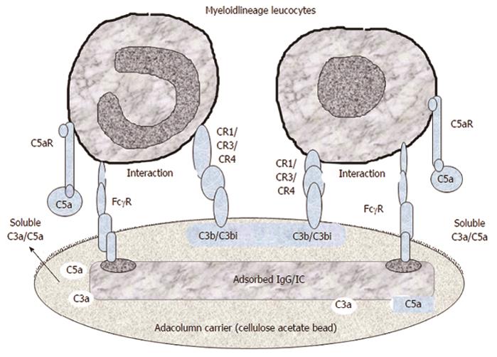
Figure 4 A tentative illustration of the mechanisms, which mediate the selective adhesion of myeloid lineage leucocytes to the granulocyte and monocyte apheresis cellulose acetate carriers.
The first event is adhesion of plasma immunoglobulin (IgG) and immune complexes (IC) to the carriers, which serve as the binding sites for the fragment crystallizable gamma receptor (FcγR) on myeloid leucocytes. Then complement activation (helped by IgG and IC) generates C3a, C5a, C3b/C3bi fragments. Of these C3b/C3bi (opsonins) adsorb onto the carriers and serve as the binding sites for the complement receptors (CR) on myeloid leucocytes. Adhesion of leucocytes results in the release of interleukin-1 receptor antagonis, hepatocyte growth factor, soluble tumour necrosis factor receptors, all with therapeutic effects (Figure 13). Modified from Reference[23].
- Citation: Saniabadi AR, Tanaka T, Ohmori T, Sawada K, Yamamoto T, Hanai H. Treating inflammatory bowel disease by adsorptive leucocytapheresis: A desire to treat without drugs. World J Gastroenterol 2014; 20(29): 9699-9715
- URL: https://www.wjgnet.com/1007-9327/full/v20/i29/9699.htm
- DOI: https://dx.doi.org/10.3748/wjg.v20.i29.9699


