Published online Apr 28, 2014. doi: 10.3748/wjg.v20.i16.4827
Revised: January 20, 2014
Accepted: March 4, 2014
Published online: April 28, 2014
Processing time: 132 Days and 16.7 Hours
The wide use of surgical endoclips in laparoscopic surgery has led to a variety of complications. Post-cholecystectomy endoclips migrating into the common bile duct after laparoscopic cholecystectomy is rare. A migrated endoclip can cause obstruction, serve as a nidus for stone formation, and cause cholangitis. While the exact pathogenesis is still unknown, it is probably related to improper clip application, subclinical bile leak, inflammation, and subsequent necrosis, allowing the clips to erode directly into the common bile duct. We present a case of endoclip migrating into the common bile duct and duodenum, resulting in choledochoduodenal fistula after laparoscopic cholecystectomy and a successful reconstruction of the biliary tract by a hepaticojejunostomy with a Roux-en-Y procedure. This case shows that surgical endoclips can penetrate into the intact bile duct wall through serial maceration, and it is believed that careful application of clips may be the only way to prevent their migration after laparoscopic cholecystectomy.
Core tip: Choledochoduodenal fistula caused by endoclip migration; an extremely rare complication after the introduction of laparoscopic cholecystectomy which can occur from days to years after laparoscopic cholecystectomy.
- Citation: Hong T, Xu XQ, He XD, Qu Q, Li BL, Zheng CJ. Choledochoduodenal fistula caused by migration of endoclip after laparoscopic cholecystectomy. World J Gastroenterol 2014; 20(16): 4827-4829
- URL: https://www.wjgnet.com/1007-9327/full/v20/i16/4827.htm
- DOI: https://dx.doi.org/10.3748/wjg.v20.i16.4827
Since the introduction of the laparoscopic technique, laparoscopic cholecystectomy is considered the gold standard for the management of symptomatic disease with a less than 3% overall complication rate[1]. Most abnormal biliary-enteric communications are the result of perforation caused by gallstones from the gallbladder or common bile duct into the duodenum, with the remainder being the result of peptic ulcer, tumor, trauma, or other local abnormalities[2] which often occur before laparoscopic cholecystectomy. Choledochoduodenal fistula caused by endoclip migration is an extremely rare complication after the introduction of laparoscopic cholecystectomy, and can occur from days to years after the procedure. We present a rare case of an endoclip migrating into the common bile duct and duodenum, resulting in choledochoduodenal fistula after the laparoscopic cholecystectomy 10 years prior.
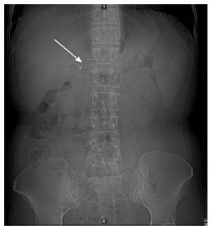
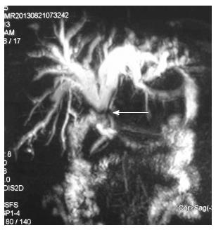
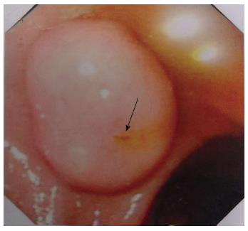
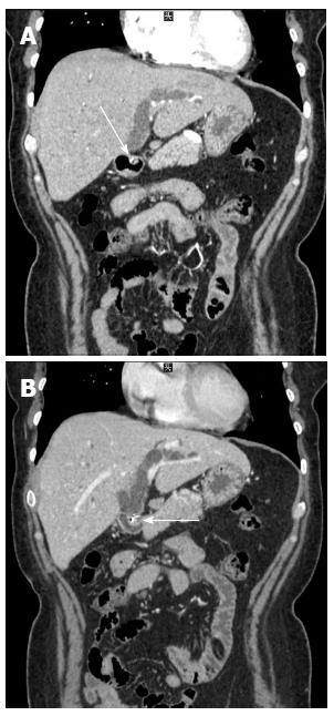
A 48-year-old woman was referred to our hospital with the chief complaint of intermittent epigastric pain, fever, and jaundice for about 3 mo. The patient underwent laparoscopic cholecystectomy (LC) 10 years previously without any intraoperative or postoperative complications. She was diagnosed as suffering acute cholangitis at a rural community hospital, with all symptoms being relieved after one week of anti-infection treatment. Physical examination at admission revealed no fever, icteric sclera, or jaundice. There was no tenderness at the epigastric area. Laboratory tests revealed white blood cells of 5470/mm3, and elevated levels of alanine aminotransferase (59 U/L, 5-40), gamma glutamyl aminotransferase (300 U/L, 0-50), and total/direct bilirubin (15.1/9.0 μmol/L, 1.7-22.5/0.0-6.0 μmol/L). Tumor markers showed high levels of CA19-9 (326 U/mL), but the levels of carcinoembryonic antigen and alpha-fetoprotein were within the normal range. A plain abdominal radiograph showed metal endoclips in the right upper quadrant area (Figure 1). Magnetic resonance imaging showed marked dilatation of the biliary duct and stenosis of the common bile duct at the hepatic duct confluence, which was close to the duodenum (Figure 2). An endoscopic image of the duodenum (Figure 3) showed yellowish bile acid leaking from a papillary orifice at the first part of duodenum wall. Computed tomography (CT) showed a mass on the duodenal wall, and linear, highly dense lesions both in the mass (Figure 4A) and in the hepatic duct confluence (Figure 4B) with dilated hepatic ducts. The patient’s clinical manifestation and imaging studies revealed a choledochoduodenal fistula caused by an injury to the common bile duct by a migrated metal endoclip. Partial resection of the common bile duct and fistula, as well as repair of the duodenum, were performed, followed by reconstruction of the biliary tract by a hepaticojejunostomy with a Roux-en-Y procedure. An endoclip was found in the duodenal portion of the choledochoduodenal fistula.
Surgical endoclips are widely used during LC as substitute ligation materials. Raoul et al[1] first reported the migration of surgical endoclips into the biliary tract acting as a nidus for stone formation after laparoscopic cholecystectomy. A variety of endoclip related complications, such as biliary leaks, endoclip migration into the common bile duct with stone formation, acute pancreatitis, cholangitis, benign stricture, obstructive jaundice, and endoclip embolism have been reported[3]. Choledochoduodenal fistula is even rarer. Biliary-enteric fistula is a known complication of chronic gallbladder disease which has a reported incidence of 0.06%-0.14%[4]. However, they usually happen before cholecystectomies, and there are no accurate data for the biliary-enteric fistula, especially for the choledochoduodenal fistula. To the best of our knowledge, this is the first report on a choledochoduodenal fistula caused by an endoclip migrating into the common bile duct and duodenum after LC.
With regard to the pathogenesis of endoclips migration after laparoscopic cholecystectomy, the first possibility is an incomplete closure of the cyst duct caused by an ineffective clip, which then brought on biloma with bile leakage. The second possibility is erosion of the bile duct wall or adjacent adhered duodenal or colonic wall because of localized inflammation around the endoclips. The eroded and inflamed common bile duct and duodenal or colonic wall would develop perforation or scar constriction, resulting in choledochoduodenal fistula or bile duct stenosis[5].
For the evaluation of choledochoduodenal fistula after LC, magnetic resonance cholangiography, endoscopic retrograde cholangiography, or CT with three-dimensional reconstruction of the biliary tract could be helpful. For the complicated structure around the fistula caused by tissue inflammation and adherence, open surgery is a safe option for reconstructing the biliary tract and repairing the defect in the duodenum due to defects in both the common bile duct and duodenum. Endoclip migration could be potentially avoided by the use of absorbable endoclips, or alternatively ultrasonic dissection without clipping[6].
In conclusion, we offer a rare case of an endoclip migrating into the common bile duct and duodenum, resulting in choledochoduodenal fistula after LC. This situation can be managed by reconstructing the biliary tract via a hepaticojejunostomy with a Roux-en-Y procedure, and could be potentially avoided by using absorbable endoclips or performing ultrasonic dissection without clipping.
Choledochoduodenal fistula caused by migration of an endoclip after laparoscopic cholecystectomy.
It should be considered in the differential diagnosis of patients with obstructive jaundice or cholangitis after laparoscopic cholecystectomy.
Diagnostic imaging must include magnetic resonance cholangiography, endoscopic retrograde cholangiography, or 3D-computed tomography with reconstruction of the biliary tract.
Surgical intervention is mostly required to reconstruct the biliary tract and to repair the defect in the duodenum due to defects in both the common bile duct and duodenum.
This is an interesting case report owing to its rarity.
P- Reviewers: La Torre F, Singhal V, Uen YH S- Editor: Zhai HH L- Editor: Rutherford A E- Editor: Ma S
| 1. | Raoul JL, Bretagne JF, Siproudhis L, Heresbach D, Campion JP, Gosselin M. Cystic duct clip migration into the common bile duct: a complication of laparoscopic cholecystectomy treated by endoscopic biliary sphincterotomy. Gastrointest Endosc. 1992;38:608-611. [RCA] [PubMed] [DOI] [Full Text] [Cited by in Crossref: 36] [Cited by in RCA: 31] [Article Influence: 0.9] [Reference Citation Analysis (0)] |
| 2. | Feller ER, Warshaw AL, Schapiro RH. Observations on management of choledochoduodenal fistula due to penetrating peptic ulcer. Gastroenterology. 1980;78:126-131. [PubMed] |
| 3. | Chong VH, Chong CF. Biliary complications secondary to post-cholecystectomy clip migration: a review of 69 cases. J Gastrointest Surg. 2010;14:688-696. [RCA] [PubMed] [DOI] [Full Text] [Cited by in Crossref: 71] [Cited by in RCA: 79] [Article Influence: 5.3] [Reference Citation Analysis (0)] |
| 4. | Lee JH, Han HS, Min SK, Lee HK. Laparoscopic repair of various types of biliary-enteric fistula: three cases. Surg Endosc. 2004;18:349. [RCA] [PubMed] [DOI] [Full Text] [Cited by in Crossref: 17] [Cited by in RCA: 13] [Article Influence: 0.6] [Reference Citation Analysis (0)] |
| 5. | Matsumoto H, Ikeda E, Mitsunaga S, Naitoh M, Furutani S, Nawa S. Choledochal stenosis and lithiasis caused by penetration and migration of surgical metal clips. J Hepatobiliary Pancreat Surg. 2000;7:603-605. [RCA] [PubMed] [DOI] [Full Text] [Cited by in Crossref: 21] [Cited by in RCA: 23] [Article Influence: 1.0] [Reference Citation Analysis (0)] |


