Copyright
©The Author(s) 1996.
World J Gastroenterol. Mar 25, 1996; 2(1): 44-50
Published online Mar 25, 1996. doi: 10.3748/wjg.v2.i1.44
Published online Mar 25, 1996. doi: 10.3748/wjg.v2.i1.44
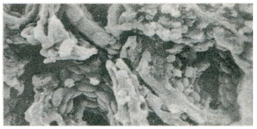
Figure 3 Focal atrophic inflammation, with a large area of cells with maintained contour but with ruptured cytomembranes and exposed cytoplasmic granules.
There were only a few red blood cells present.
- Citation: Guang-Yao Y, Fen-Xue H, Fen YY. Clinical and experimental study on gastric mucosal pathology, gene expression, cAMP and trace elements of pixu patients. World J Gastroenterol 1996; 2(1): 44-50
- URL: https://www.wjgnet.com/1007-9327/full/v2/i1/44.htm
- DOI: https://dx.doi.org/10.3748/wjg.v2.i1.44


