Published online Sep 28, 2013. doi: 10.3748/wjg.v19.i36.6108
Revised: July 10, 2013
Accepted: July 17, 2013
Published online: September 28, 2013
Processing time: 128 Days and 6 Hours
A 46-year-old man was admitted with obstructive jaundice and cross-sectional imaging with computed tomography suggested distal biliary obstruction. A distal common bile duct stricture was found at endoscopic retrograde cholangiopancreatography (ERCP) and cytology was benign. A 6 cm fully covered self-expanding metal stent (SEMS) was inserted across the stricture to optimize biliary drainage. However, the SEMS could not be removed at repeat ERCP a few months later. A further fully covered SEMS was inserted within the existing stent to enable extraction and both stents were retrieved successfully a few weeks later. Fully covered biliary (SEMS) are used to treat benign biliary strictures. This is the first reported case of inability to remove a fully-covered biliary SEMS. Possible reasons for this include tissue hyperplasia and consequent overgrowth into the stent proximally, or chemical or mechanical damage to the polymer covering of the stent. Application of the stent-in-stent technique allowed successful retrieval of the initial stent.
Core tip: Inability to retrieve a fully covered biliary self-expanding metal stent (SEMS) due to potential fixation of the stent to the duct wall can be addressed by the insertion of a second SEMS within the existing one, which facilitates the release of the initial SEMS.
- Citation: Menon S. Removal of an embedded "covered" biliary stent by the "stent-in-stent" technique. World J Gastroenterol 2013; 19(36): 6108-6109
- URL: https://www.wjgnet.com/1007-9327/full/v19/i36/6108.htm
- DOI: https://dx.doi.org/10.3748/wjg.v19.i36.6108
The endoscopic management of benign biliary strictures has been transformed by the advent of removable fully covered metal stents. We report a case in which a fully covered biliary stent could not be removed endoscopically initially.
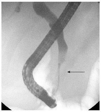
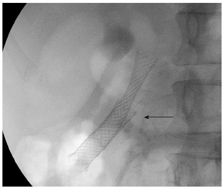
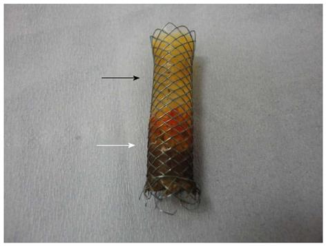
A 46-year-old man was admitted with abdominal pain and obstructive jaundice [Bilirubin 14 mg/dL, Alanine aminotransferase (ALT) 123 IU/L, Alkaline phosphatase (ALP) 640 IU/L and a raised Amylase 350 U/L]. Cross-sectional imaging with computed tomography confirmed an enlarged and markedly calcified head of pancreas (Figure 1). Endoscopic retrograde cholangiopancreatography (ERCP) revealed a 2.5 cm distal common bile duct (CBD) stricture with proximal biliary dilatation. A 6 cm fully covered self-expanding metal stent (SEMS) (Wallflex, Boston Scientific, Boston, MA, United States) was inserted across the stricture. Cytology from the stricture was benign. At repeat ERCP 5 mo later, the SEMS could not be removed endoscopically with suggestion of the stent having embedded into the bile duct wall. Mild residual stricturing was noted at the apex of the SEMS. Further dilatation of the stricture was carried out to 10 mm and an in-stent dilatation was carried out to 12 mm to release the stent from the duct wall without success. At further ERCP in the next few weeks, a second 6 cm fully covered SEMS was inserted across the existing stent (Figure 2). Six weeks later, both stents were removed easily. Cholangiography suggested complete resolution of the original stricture. Examination of the initially inserted stent suggested possible damage to the silicone polymer stent covering (Figure 3) and is subject to further investigation.
Embedded SEMS in the esophagus can be removed by inserting a fully covered SEMS within the SEMS, which causes pressure necrosis of tissue across the uncovered portion of the embedded SEMS, thereby facilitating its removal[1]. This technique has been used previously for removing uncovered biliary SEMS[2,3].
Fully covered biliary (SEMS) are used widely to treat benign biliary strictures. This is the first reported case of inability to remove a fully-covered biliary SEMS. Possible reasons for this include tissue hyperplasia and consequent overgrowth into the stent proximally, or chemical or mechanical damage to the polymer covering of the stent. Application of the stent-in-stent technique allowed successful retrieval of the initial stent.
P- Reviewer Kamisawa T S- Editor Wen LL L- Editor A E- Editor Ma S
| 1. | Hirdes MM, Siersema PD, Houben MH, Weusten BL, Vleggaar FP. Stent-in-stent technique for removal of embedded esophageal self-expanding metal stents. Am J Gastroenterol. 2011;106:286-293. [RCA] [PubMed] [DOI] [Full Text] [Cited by in Crossref: 85] [Cited by in RCA: 83] [Article Influence: 5.9] [Reference Citation Analysis (0)] |
| 2. | Arias Dachary FJ, Chioccioli C, Deprez PH. Application of the “covered-stent-in-uncovered-stent” technique for easy and safe removal of embedded biliary uncovered SEMS with tissue ingrowth. Endoscopy. 2010;42 Suppl 2:E304-E305. [RCA] [PubMed] [DOI] [Full Text] [Cited by in Crossref: 16] [Cited by in RCA: 17] [Article Influence: 1.2] [Reference Citation Analysis (0)] |
| 3. | Tan DM, Lillemoe KD, Fogel EL. A new technique for endoscopic removal of uncovered biliary self-expandable metal stents: stent-in-stent technique with a fully covered biliary stent. Gastrointest Endosc. 2012;75:923-925. [RCA] [PubMed] [DOI] [Full Text] [Cited by in Crossref: 19] [Cited by in RCA: 20] [Article Influence: 1.5] [Reference Citation Analysis (0)] |


