Published online Apr 14, 2013. doi: 10.3748/wjg.v19.i14.2282
Revised: January 16, 2013
Accepted: January 23, 2013
Published online: April 14, 2013
Processing time: 115 Days and 8.8 Hours
Sarcina ventriculi is a Gram positive organism, which has been reported to be found rarely, in the gastric specimens of patients with gastroparesis. Only eight cases of Sarcina, isolated from gastric specimens have been reported so far. Sarcina has been implicated in the development of gastric ulcers, emphysematous gastritis and gastric perforation. We report a case of 73-year-old male, with history of prior Billroth II surgery and truncal vagotomy, who presented for further evaluation of iron deficiency anemia. An upper endoscopy revealed diffuse gastric erythema, along with retained food. Biopsies revealed marked inflammation with ulcer bed formation and presence of Sarcina organisms. The patient was treated with ciprofloxacin and metronidazole for 1 wk, and a repeat endoscopy showed improvement of erythema, along with clearance of Sarcina organisms. Review of reported cases including ours suggests that Sarcina is more frequently an innocent bystander rather than a pathogenic organism. However, given its association with life threatening illness in two reported cases, it may be prudent to treat with antibiotics and anti-ulcer therapy, until further understanding is achieved.
Core tip:Sarcina ventriculi is a rare bacterium, seen in gastric biopsies of patients with gastroparesis. Only eight cases have been reported so far, where in it has been implicated in the development of gastric ulcers, emphysematous gastritis and gastric perforation. In our case, gastric erythema improved with antibiotic treatment. Given its association with life threatening illness in two reported cases, it may be prudent to treat with antibiotics and anti-ulcer therapy, until further understanding is achieved.
-
Citation: Ratuapli SK, Lam-Himlin DM, Heigh RI.
Sarcina ventriculi of the stomach: A case report. World J Gastroenterol 2013; 19(14): 2282-2285 - URL: https://www.wjgnet.com/1007-9327/full/v19/i14/2282.htm
- DOI: https://dx.doi.org/10.3748/wjg.v19.i14.2282
Sarcina ventriculi is a Gram positive anaerobic bacterium, with carbohydrate fermentative metabolism as its sole energy source[1], and is able survive in very low pH environment[2]. Even though it is similar in appearance to Micrococcus species, certain morphological features (i.e., larger size, non-cluster forming pattern) help differentiate it from the latter organism[3].
Various reports in veterinary literature have implicated Sarcina in the development of gastric dilatation[4] and death of livestock, cats and horses[5,6]. Sarcina has also been reported to be found in feces of healthy humans consuming a predominantly vegetarian diet[7]. Recently, several reports have shown an association between Sarcina in the stomach and chronic nausea, dyspepsia, abdominal pain, gastric ulcers[3], and rarely emphysematous gastritis[8] and gastric perforation[9]. However Sarcina has also been found in gastric specimens without any other pathologic changes[3], suggesting that it may be a bystander rather than a pathogenic organism. To date, only eight cases of Sarcina ventriculi isolated from gastric biopsy specimens have been reported. We now report a case of Sarcina ventriculi of the stomach, associated with iron deficiency anemia and gastroparesis.
A 73-year-old male presented to the clinic for further evaluation of iron deficiency anemia. The patient had a history of medically refractory gastric ulcers in his 20 s, for which he underwent antrectomy and gastrojejunostomy (Billroth II) along with truncal vagotomy in 1985. He continued to be anemic since the surgery, with intermittent intake of oral iron replacement.
On initial evaluation for an incidentally detected anemia prior to unrelated urologic surgery, he did not have any gastrointestinal symptoms. The patient specifically denied nausea, vomiting, abdominal pain or weight loss. His complete blood count revealed decreased hemoglobin of 8.5 g/dL, decreased mean corpuscular volume of 63.2 fL, normal white cell count of 9.8 × 10⁹ L (normal 4.2 × 10⁹-10.2 × 10⁹ L) and elevated platelet count 415 × 109 L (normal 151 × 109-355 × 109 L). Iron studies showed markedly reduced iron level of 12 mg/dL (normal 50-150 mg/dL), with an elevated total iron binding capacity of 490 mg/dL (normal 250-400 mg/dL) and reduced iron % saturation of 2% (normal 14%-50%).
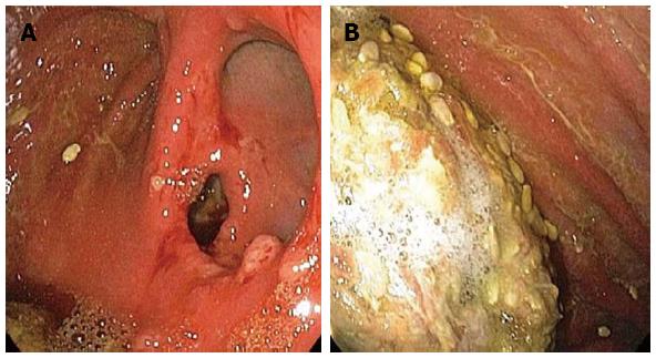
Three years prior, an esophagogastroduodenoscopy (EGD) had revealed an anastomotic ulcer and polyps at the anastomotic site, the biopsies of which showed acute inflammation, but were otherwise unremarkable. A colonoscopy at that time was unremarkable except for diverticulosis. An EGD done during the current evaluation demonstrated diffuse gastric erythema, along with two 4mm polyps at the anastomosis (Figure 1). There was also a large amount of retained food in the stomach.
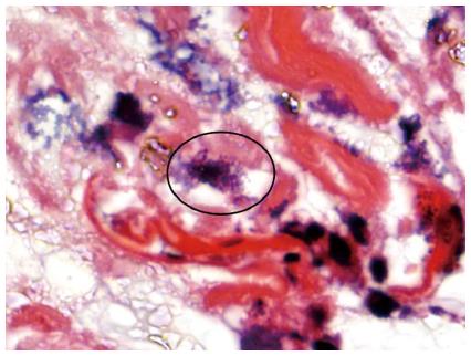
Tissue biopsies of the erythematous stomach revealed marked inflammation with ulcer bed formation, along with abundant bacterial overgrowth including the presence of Sarcina organisms (Figure 2). The Sarcina organisms were identified on routine hematoxylin and eosin (HE) stain, and no additional special stains or immunolabeling was performed. Based on prior studies[3], the tetrad morphology and size are characteristic enough to establish a diagnosis without further ancillary testing. Biopsies were negative for Helicobacter pylori, both by routine HE staining and by immunohistochemical staining. Aspirates from the small bowel also came back positive for small intestinal bacterial overgrowth, with > 100 000 cfu/mL of mixed Gram-positive and Gram-negative flora.
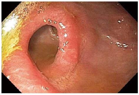
The patient was treated with metronidazole 250 mg three times a day and ciprofloxacin 250 mg twice daily for 1 wk, along with daily sucralfate. He also received intravenous (IV) iron 300 mg × 2 doses followed by oral iron and achieved normal iron stores and hemoglobin levels. Subsequent follow up with a repeat EGD 3 mo later showed improvement of gastric erythema, and absence of food bezoar (Figure 3). Aspirates from the small bowel continued to suggest small intestinal bacterial overgrowth, with > 100 000 cfu/mL. However, repeat biopsies from the stomach were negative for Sarcina organisms, and showed features of chronic gastritis. Clinically, the patient’s perception of overall health improved with the above treatment, and he continued to be free of gastrointestinal symptoms.
While the pathogenic role of Sarcina in the veterinary literature is well established, its role in human disease is not entirely clear. Since the initial description in 1842, the pathogenic role in humans has been questioned, as it has been found in the blood[10] and feces[7] of healthy humans.
Over the last three years, 8 cases[3,8,9] of Sarcina associated with endoscopic biopsies have been reported. While all these patients presented with various gastrointestinal symptoms (nausea, vomiting, epigastric pain, dyspepsia) only two patients had associated life threatening complications of emphysematous gastritis[8] and gastric perforation[9] (Table 1). Our patient did not have any gastrointestinal symptoms, and Sarcina was found incidentally, when gastric biopsies were performed for erythematous mucosa.
| Case No. | Age | Sex | Symptoms/clinical findings | Endoscopic findings | Histologic findings | Treatment | Follow-up |
| 1 | 14 | Male | Abdominal pain | Not performed | Diffuse acute hemorrhagic gastritis and Sarcina organisms | Gentamicin and metronidazole | Symptoms improved after 5 d and patient discharged |
| CT showed pneumoperitoneum. | |||||||
| Intraoperatively there was necrotic stomach and gastric perforation and peritonitis | |||||||
| 2 | 50 | Male | Chronic nausea, vomiting | Esophagitis, duodenal lesion | Chronic superficial gastritis and ulcer with Sarcina organisms | Not available | Not available |
| 3 | 3 | Female | Vomiting, hematemesis Abdominal X-ray showed dilated stomach with intramural air | Gastric inflammation, blackening of mucosa, cobblestone appearance | Polymorphic inflammatory infiltrate with Sarcina organisms and gas bubbles | Imipene, fluconazole and omeprazole | Repeat endoscopy 6 mo later showed complete normalization |
| 4 | 58 | Female | Nausea and vomiting | Gastritis, food bezoar, inflammatory mass in duodenum | Active chronic gastritis with Sarcina organisms | Partial gastrectomy for obstruction | Treated for adenocarcinoma of pylorus |
| 5 | 44 | Female | Dyspepsia and substernal burning | Gastric ulcer and retained food | Non malignant gastric ulcer with Sarcina organisms | Omeprazole, ranitidine, metoclopramide | Symptoms improved |
| 6 | 36 | Male | Nausea, vomiting, epigastric pain in the setting of narcotic use | Retained food | Sarcina organisms without other histologic abnormalities | Received jejunostomy for malnutrition | Repeat biopsy negative for Sarcina organisms |
| 7 | 12 | Female | Dysphagia in the setting of esophageal atresia status post gastric pull through | Retained food, anastamotic stricture | Reflux esophagitis, Sarcina organisms | Information unavailable | Information unavailable |
| 8 | 46 | Female | Epigastric pain in the setting of pancreatic adenocarcinoma status post pancreatico-duodenectomy | Retained food and bile | Active chronic duodenitis with Sarcina organisms | No treatment | Continues spasms after 1 mo |
Another interesting feature is the presence of delayed gastric emptying in five of the eight reported cases. All of these patients had retained food in stomach during endoscopic examination. Similarly, our patient had a Billroth II with truncal vagotomy, which predisposed him to have delayed gastric emptying, as was evident by the gastric bezoar seen during endoscopic examination. Hence impaired emptying of stomach could potentially lead to the growth of Sarcina in the stomach.
The need for antibiotic treatment, when Sarcina is found in endoscopic biopsies of clinically stable patients is unknown. Of the reported cases, two patients with associated life threatening disease (i.e., emphysematous gastritis and gastric perforation) received intravenous antibiotics and recovered. One patient with non-life threatening disease was treated with combination of proton pump inhibitors and prokinetics, with good relief of symptoms. Some authors suggest that an underlying mucosal defect, such as erosion or ulceration, may predispose patients to more serious sequelae, from this otherwise ubiquitous organism[3]. We elected to treat our patient with antibiotics, as there was significant gastric erythema and ulceration, as well as small intestinal bacterial overgrowth.
In summary, Sarcina ventriculi is a rare bacterium, seen predominantly in patients with delayed gastric emptying. Review of the published cases along with our case suggests that it is more frequently an innocent bystander rather than a pathogenic organism. Given its association with life threatening illness in two reported cases, it may be prudent to treat with antibiotics and anti-ulcer therapy, until further understanding is achieved.
P- Reviewer Fujiya M S- Editor Gou SX L- Editor A E- Editor Zhang DN
| 1. | Claus D, Wilmanns H. Enrichment and selective isolation of Sarcina maxima Lindner. Arch Microbiol. 1974;96:201-204. [RCA] [PubMed] [DOI] [Full Text] [Cited by in Crossref: 8] [Cited by in RCA: 9] [Article Influence: 0.2] [Reference Citation Analysis (0)] |
| 2. | Lowe SE, Pankratz HS, Zeikus JG. Influence of pH extremes on sporulation and ultrastructure of Sarcina ventriculi. J Bacteriol. 1989;171:3775-3781. [PubMed] |
| 3. | Lam-Himlin D, Tsiatis AC, Montgomery E, Pai RK, Brown JA, Razavi M, Lamps L, Eshleman JR, Bhagavan B, Anders RA. Sarcina organisms in the gastrointestinal tract: a clinicopathologic and molecular study. Am J Surg Pathol. 2011;35:1700-1705. [RCA] [PubMed] [DOI] [Full Text] [Cited by in Crossref: 57] [Cited by in RCA: 82] [Article Influence: 5.9] [Reference Citation Analysis (0)] |
| 4. | Edwards GT, Woodger NG, Barlow AM, Bell SJ, Harwood DG, Otter A, Wight AR. Sarcina-like bacteria associated with bloat in young lambs and calves. Vet Rec. 2008;163:391-393. [PubMed] |
| 5. | DeBey BM, Blanchard PC, Durfee PT. Abomasal bloat associated with Sarcina-like bacteria in goat kids. J Am Vet Med Assoc. 1996;209:1468-1469. [PubMed] |
| 6. | Vatn S, Gunnes G, Nybø K, Juul HM. Possible involvement of Sarcina ventriculi in canine and equine acute gastric dilatation. Acta Vet Scand. 2000;41:333-337. [PubMed] |
| 7. | Crowther JS. Sarcina ventriculi in human faeces. J Med Microbiol. 1971;4:343-350. [RCA] [PubMed] [DOI] [Full Text] [Cited by in Crossref: 41] [Cited by in RCA: 48] [Article Influence: 0.9] [Reference Citation Analysis (0)] |
| 8. | Laass MW, Pargac N, Fischer R, Bernhardt H, Knoke M, Henker J. Emphysematous gastritis caused by Sarcina ventriculi. Gastrointest Endosc. 2010;72:1101-1103. [PubMed] |
| 9. | Tolentino LE, Kallichanda N, Javier B, Yoshimori R, French SW. A case report of gastric perforation and peritonitis associated with opportunistic infection by Sarcina ventriculi. Lab Med. 2003;34:535-537. [RCA] [DOI] [Full Text] [Cited by in Crossref: 26] [Cited by in RCA: 16] [Article Influence: 0.7] [Reference Citation Analysis (0)] |
| 10. | Ferrier D. The Constant Occurrence of Sarcina Ventriculi (Goodsir) in the Blood of Man and the Lower Animals: With Remarks on the Nature of Sarcinous Vomiting. Br Med J. 1872;1:98-99. [RCA] [PubMed] [DOI] [Full Text] [Cited by in Crossref: 16] [Cited by in RCA: 20] [Article Influence: 1.4] [Reference Citation Analysis (0)] |


