Published online Nov 21, 2012. doi: 10.3748/wjg.v18.i43.6341
Revised: August 14, 2012
Accepted: August 25, 2012
Published online: November 21, 2012
An isolated parenchymal gastric metastasis from ovarian carcinoma without any other sites of recurrence is extremely rare. Only two cases have been reported, both of which were symptomatic. We herein report such a case without any symptoms. A 61-year-old woman presented with a high cancer antigen-125 level without any other clinical manifestation. A subsequent 18F-fluorodeoxyglucose (18F-FDG) positron emission tomography/computed tomography scan revealed a submucosal mass with hypermetabolism of 18F-FDG (standardized uptake value: 5.36) in the gastric antrum. The final pathology after gastric antrectomy showed a metastatic gastric tumor from a primary ovarian carcinoma. We also performed an extensive literature review about gastric metastasis from ovarian carcinoma published until recently, and this is the first case of an isolated parenchymal gastric metastasis from ovarian carcinoma without any symptoms.
- Citation: Zhou JJ, Miao XY. Gastric metastasis from ovarian carcinoma: A case report and literature review. World J Gastroenterol 2012; 18(43): 6341-6344
- URL: https://www.wjgnet.com/1007-9327/full/v18/i43/6341.htm
- DOI: https://dx.doi.org/10.3748/wjg.v18.i43.6341
Ovarian carcinoma usually metastasizes along the peritoneum throughout the pelvic and abdominal cavity, such as pelvic wall, omentum and mesentery. Gastrointestinal involvement is not common. Even it happens, gastrointestinal tract metastasis of ovarian carcinoma is merely limited to serosa. Solitary parenchymal gastric metastasis from ovarian carcinoma is extremely rare, and only two cases have been reported in English up till now[1,2]. We herein present a case of gastric metastasis from ovarian carcinoma without any symptoms and other sites of recurrence.
In December 2011, a 61-year-old woman was admitted to our hospital because of a high cancer antigen (CA)-125 level of up to 116.5 U/mL (normal, < 35U/mL), and she had no epigastric pain and fullness, hematemesis, melena, weight loss and any other clinical manifestations. In 1999, she underwent optimal debulking cytoreductive surgery in our hospital for ovarian adenocarcinoma, followed by ten cycles of adjuvant chemotherapy with cisplatin and cyclophosphamide. In May 2006, when her CA-125 level increased to 57.9 U/mL, she received another ten cycles of adjuvant chemotherapy with taxol, cyclophosphamide, carboplatin and bleomycin. CA-125 level was tested every two months and it exceeded the normal range again in December 2011.
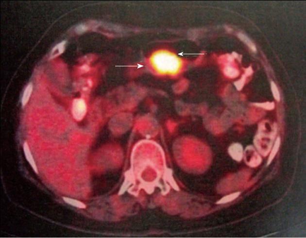
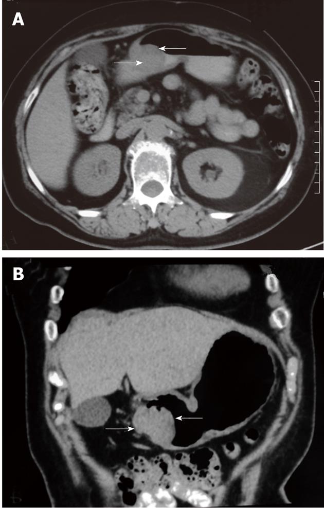
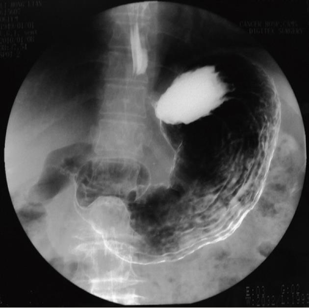
18F-fluorodeoxyglucose positron emission tomography/computed tomography (18F-FDG PET/CT) scanning for ruling out the recurrent ovarian carcinoma that was suspected due to the CA-125 level. 18F-FDG PET/CT revealed a mass located in gastric antrum with high 18F-FDG uptake (standardized uptake value: 5.36) (Figure 1), and there were no any other lesions with high 18F-FDG uptake in the abdominopelvic region. A subsequent non-contrast-enhanced CT displayed a 2.4 cm × 3.0 cm submucosal mass in the gastric antrum (Figure 2), which had not been found in the CT scanning done on April 4, 2010. The patient could not tolerate and refuse to take endoscopic examination, so we performed gastroenterography instead. Upper gastroenterography also showed clearly a lesion with a tiny ulceration on the surface of gastric mucosa (Figure 3).
The patient then underwent local gastrectomy. During the operation, we found that both mucosa and serosa were involved, but there was no intumescent lymph node around the gastric antrum. The incision of gastric antrum was fixed with a double-layer hand-sewn suture transversely.
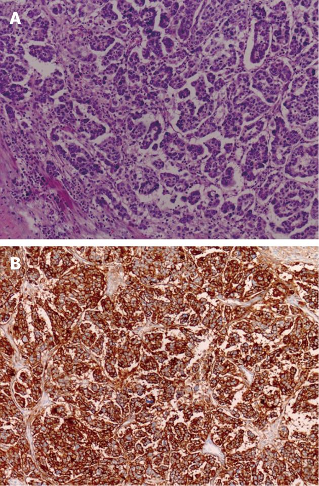
On cut section, a gray-white tumor of 3.2 cm × 2.8 cm × 3.5 cm was situated in the muscularis propria and bulged into the serosa. Microscopically (Figure 4A), serous papillary adenocarcinoma cells infiltrated into normal gastric tissues with cancer embolus in the vessels. There was a deep ulceration on the overlying mucosa. A non-metastatic lymph node was found in the specimen. Values of the immunohistochemical detection of the tumor cells (Figure 4B) were: CA-125 (+++), Wilms’ tumor-1 (+++), estrogen receptor (++), cytokeratin 7(+), cytokeratin 20(-), progesterone receptor (-) and CDX-2 (-). The immunohistochemical staining result supported the final diagnosis of gastric metastasis from ovarian serous adenocarcinoma.
CA-125 level was decreased to 53.1 U/mL on the 7th postoperative day. Her postoperative course was unremarkable and she was discharged on the 9th day after operation. When this manuscript was submitted, she had no experience of recurrent disease.
Metastatic disease involving stomach is unusual. A study found 17 metastases to the stomach among 1010 patients with malignant tumors, giving a frequency of 1.7%[3]. Another series of autopsies discovered 92 gastric metastases among 7165 cases, with a rate of 1.28%[4]. Most gastric metastases arise from primary breast cancer, followed by melanoma and lung cancer. The incidence of gastric metastases was 3.6% (25/694) in patients with breast cancer and 1.3% (10/747) in patients with lung cancer. No study had analyzed the incidence of gastric metastasis from ovarian carcinoma due to the extremely rare occurrence. According to our review of the literature, there has been no report of gastric metastases from ovarian carcinoma in Chinese.
We performed a very comprehensive review of all case reports of gastric metastasis from ovarian carcinoma. Until this April, ten other reports (Table 1) in English could be searched in PubMed. Patient age ranged from 42 years to 70 years. Two cases[10,12] were diagnosed with primary ovarian carcinoma simultaneously, the longest time from diagnosis of primary tumor to discovery of gastric metastasis being 18 years[2]. Clinical manifestations were diversified and nonspecific, and three cases were asymptomatic (3/11, 27.27%).
| Author | Age | Histology | Recurrence sites | Recurrence time | Symptoms | Survival |
| Sangha et al[1] | 55 | NR | Stomach | 7 yr | Belching reflux, epigastic discomfort | NED NR |
| Pernice et al[2] | 42 | Adenocarcinoma G3 | Stomach + perigastric area | 18 yr | Asymptomatic | 12 mo NED |
| Taylor et al[5] | 62 | Serous adenocarcinoma G3 | Lung + liver + stomach | 10 mo | Haemorragy | 6 mo DOD |
| Kobayashi et al[6] | 48 | NR | Spleen + pancreas+ sigmoid colon | 21 yr | Hemorrhage, partial bowel obstruction | NR |
| Dupuychaffay et al[7] | 65 | Adenocarcinoma G3 | Stomach + diaphragm + pancreas + peritoneal nodes | 16 yr | Fever, aasthenia, anorexia, epigastric pain | NR |
| Bechade et al[8] | 51 | Adenocarcinoma G3 | Stomach + peritoneal nodes + ovaries | NR | Hemorrhage | ED NR |
| Jung et al[9] | 49 | Serous ovarian adenocarcinoma | Gastric antrum + presacral area | 52 mo | Asymptomatic | 18 mo NED |
| Carrara et al[10] | 70 | Adenocarcinoma | Gastric body | Simultaneouly | Mild anemia, dyspepsia | NR |
| Majeurs et al[11] | 61 | Serous adenocarcinoma G3 | Stomach + sigmoid colon | 7 mo | Epigastic discomfort, vomit | 18 mo DOD |
| Kang et all[12] | 55 | Adenocarcinoma | Gastric antrum + pelvic cavity | Simultaneouly | Epigastric pain, abdominal distention | 12 mo NED |
| Present case | 61 | Adenocarcinoma G3 | Gastric antrum | 12 yr | Asymptomatic | 5 mo NED |
Due to the extremely low incidence, it is hard to make a correct diagnosis of gastric metastasis from ovarian carcinoma. According to our literature review, some cases[2,9] only presented with CA-125 levels beyond normal range but without any symptoms. Since CT scanning, gastroenterography and gastroscopy all showed a submucosal tumor of stomach, a wrong diagnosis of gastrointestinal stromal tumor[12] would be easily made[12]. So, when a patient has a history of ovarian carcinoma, especially when her CA-125 level is high, metastasis from ovarian carcinoma should be considered. 18F-FDG PET/CT can be useful. In our case, 18F-FDG PET/CT scanning revealed a high metabolic uptake lesion of gastric antrum, which is similar to the findings as described by other authors[2,12].
Ovarian carcinoma is more likely to metastasize along the peritoneal surface, but the mechanism of gastric metastasis remains unclear, it may be because of the rich blood supply of stomach. Local excision without radical lymphadenectomy following adjuvant chemotherapy is effective and recommended for metastases of ovarian carcinoma. The prognosis of gastric metastases of ovarian carcinoma remains unknown, according to our literature review, a one-year survival rate can be expected optimistically (5/6, 83.33%).
We want to thank our colleagues from the Department of Pathology for providing the pathological pictures.
Peer reviewers: Yasuhiro Kodera, MD, PhD, FACS, Associate Professor, Department of Surgery II, Nagoya University Graduate School of Medicine, 65 Tsurumai-cho, Showa-ku, Nagoya, Aichi 466-8550, Japan; Jan Kulig, Professor, MD, 1st Department of General and GI Surgery, Jagiellonian University Medical College, 40 Kopernika St., 31-501 Kraków, Poland
S- Editor Gou SX L- Editor A E- Editor Xiong L
| 1. | Sangha S, Gergeos F, Freter R, Paiva LL, Jacobson BC. Diagnosis of ovarian cancer metastatic to the stomach by EUS-guided FNA. Gastrointest Endosc. 2003;58:933-935. [RCA] [PubMed] [DOI] [Full Text] [Cited by in Crossref: 12] [Cited by in RCA: 17] [Article Influence: 0.8] [Reference Citation Analysis (0)] |
| 2. | Pernice M, Manci N, Marchetti C, Morano G, Boni T, Bellati F, Panici PB. Solitary gastric recurrence from ovarian carcinoma: a case report and literature review. Surg Oncol. 2006;15:267-270. [RCA] [PubMed] [DOI] [Full Text] [Cited by in Crossref: 5] [Cited by in RCA: 7] [Article Influence: 0.4] [Reference Citation Analysis (0)] |
| 3. | Menuck LS, Amberg JR. Metastatic disease involving the stomach. Am J Dig Dis. 1975;20:903-913. [RCA] [PubMed] [DOI] [Full Text] [Cited by in Crossref: 138] [Cited by in RCA: 135] [Article Influence: 2.7] [Reference Citation Analysis (0)] |
| 4. | Berge T, Lundberg S. Cancer in Malmö 1958-1969. An autopsy study. Acta Pathol Microbiol Scand Suppl. 1977;1-235. [PubMed] |
| 5. | Taylor RR, Phillips WS, O'Connor DM, Harrison CR. Unusual intramural gastric metastasis of recurrent epithelial ovarian carcinoma. Gynecol Oncol. 1994;55:152-155. [RCA] [PubMed] [DOI] [Full Text] [Cited by in Crossref: 19] [Cited by in RCA: 21] [Article Influence: 0.7] [Reference Citation Analysis (0)] |
| 6. | Kobayashi O, Sugiyama Y, Cho H, Tsuburaya A, Sairenji M, Motohashi H, Yoshikawa T. Clinical and pathological study of gastric cancer with ovarian metastasis. Int J Clin Oncol. 2003;8:67-71. [RCA] [PubMed] [DOI] [Full Text] [Cited by in Crossref: 17] [Cited by in RCA: 22] [Article Influence: 1.0] [Reference Citation Analysis (0)] |
| 7. | Dupuychaffray JP, Auger C, Funes De La Vega M, Riche A, Boulanger V, Blanchot P. [Gastric metastasis from ovarian carcinoma revealed by a gastro-splenic perforation]. Gastroenterol Clin Biol. 2004;28:490-493. [RCA] [PubMed] [DOI] [Full Text] [Cited by in Crossref: 2] [Cited by in RCA: 3] [Article Influence: 0.2] [Reference Citation Analysis (0)] |
| 8. | Béchade D, Desramé J, Raynaud JJ, Eggenspieler P, Baranger B, Védrine L, Ceccaldi B, Algayres JP. [Hemorrhagic gastric metastasis of an ovarian carcinoma]. Gastroenterol Clin Biol. 2005;29:1065-1066. [PubMed] |
| 9. | Jung HJ, Lee HY, Kim BW, Jung SM, Kim HG, Ji JS, Choi H, Lee BI. Gastric Metastasis from Ovarian Adenocarcinoma Presenting as a Submucosal Tumor without Ulceration. Gut Liver. 2009;3:211-214. [RCA] [PubMed] [DOI] [Full Text] [Full Text (PDF)] [Cited by in Crossref: 13] [Cited by in RCA: 20] [Article Influence: 1.3] [Reference Citation Analysis (0)] |
| 10. | Carrara S, Doglioni C, Arcidiacono PG, Testoni PA. Gastric metastasis from ovarian carcinoma diagnosed by EUS-FNA biopsy and elastography. Gastrointest Endosc. 2011;74:223-225. [RCA] [PubMed] [DOI] [Full Text] [Cited by in Crossref: 13] [Cited by in RCA: 13] [Article Influence: 0.9] [Reference Citation Analysis (0)] |
| 11. | Majerus B, Timmermans M. [Gastric metastases of ovarian adenocarcinoma. Apropos of a case]. Acta Chir Belg. 1990;90:166-171. [PubMed] |
| 12. | Kang WD, Kim CH, Cho MK, Kim JW, Lee JS, Ryu SY, Kim YH, Choi HS, Kim SM. Primary epithelial ovarian carcinoma with gastric metastasis mimic gastrointestinal stromal tumor. Cancer Res Treat. 2008;40:93-96. [RCA] [PubMed] [DOI] [Full Text] [Cited by in Crossref: 8] [Cited by in RCA: 14] [Article Influence: 0.8] [Reference Citation Analysis (0)] |


