Copyright
©2012 Baishideng Publishing Group Co.
World J Gastroenterol. Jan 14, 2012; 18(2): 192-196
Published online Jan 14, 2012. doi: 10.3748/wjg.v18.i2.192
Published online Jan 14, 2012. doi: 10.3748/wjg.v18.i2.192
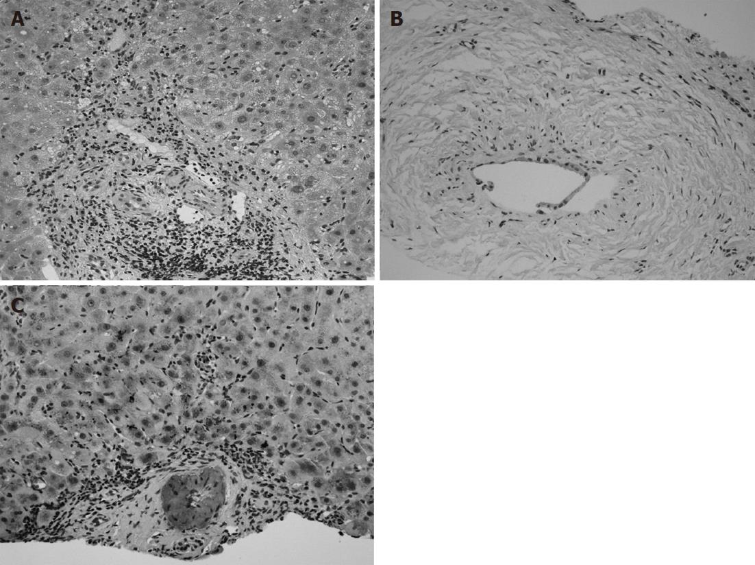
Figure 3 Histology of the liver by hematoxylin and eosin stain.
An increase in lymphoneutrophilic infiltrates in the portal tracts, interface hepatitis with ductular proliferation, cholate stasis (400 ×, A), and damaged interlobular bile ducts with collagenous periductal thickening (200 ×, B) were revealed. Amyloid deposition in the vessel walls of the portal tracts was also apparent in immunohistochemical staining (200 ×, C).
- Citation: Kato T, Komori A, Bae SK, Migita K, Ito M, Motoyoshi Y, Abiru S, Ishibashi H. Concurrent systemic AA amyloidosis can discriminate primary sclerosing cholangitis from IgG4-associated cholangitis. World J Gastroenterol 2012; 18(2): 192-196
- URL: https://www.wjgnet.com/1007-9327/full/v18/i2/192.htm
- DOI: https://dx.doi.org/10.3748/wjg.v18.i2.192


