Copyright
©2011 Baishideng Publishing Group Co.
World J Gastroenterol. Feb 7, 2011; 17(5): 666-670
Published online Feb 7, 2011. doi: 10.3748/wjg.v17.i5.666
Published online Feb 7, 2011. doi: 10.3748/wjg.v17.i5.666
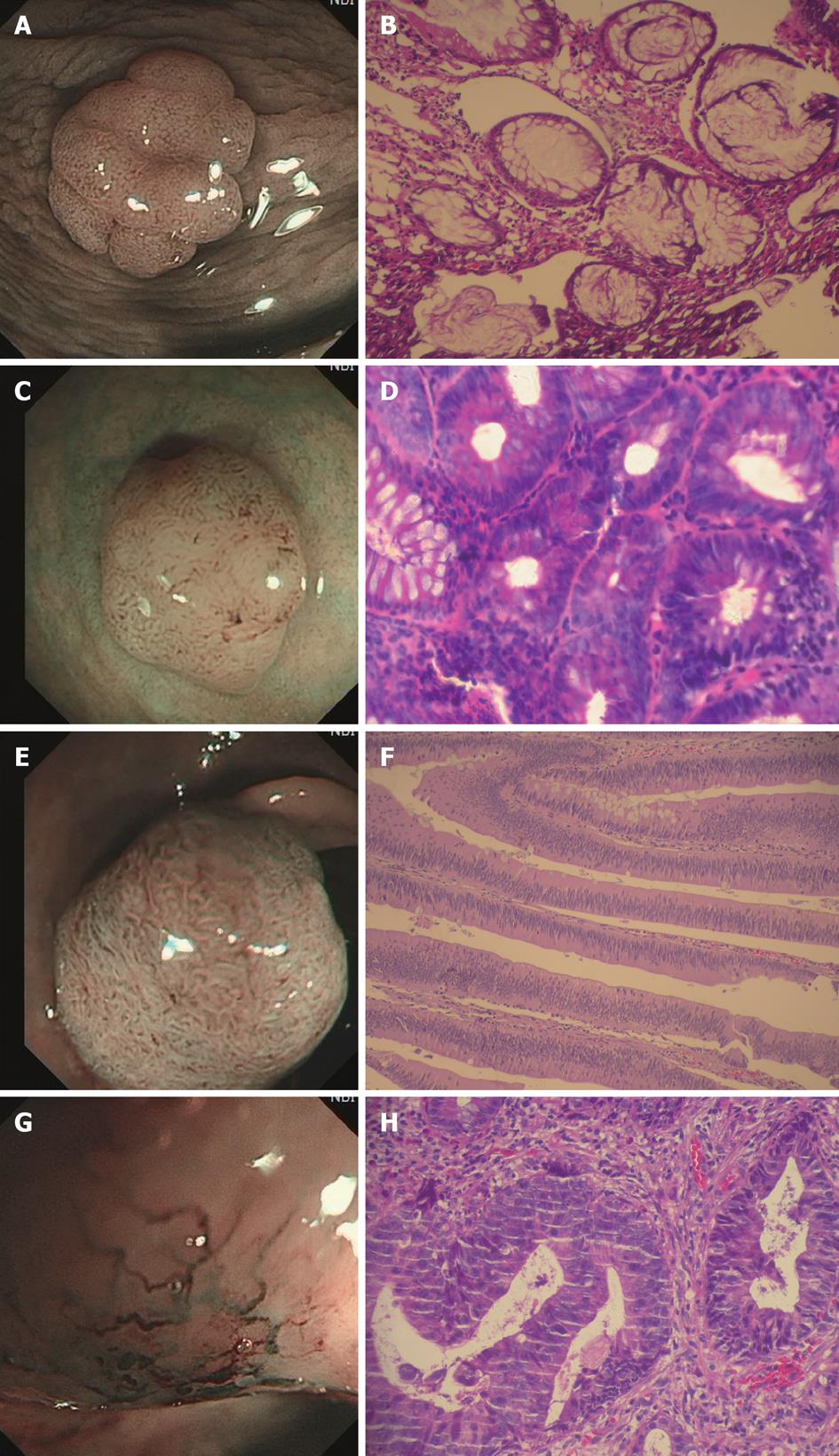
Figure 1 Narrow-band imaging endoscopic evaluation and the corresponding pathological images.
A, B: A hyperplastic polyp with narrow-band imaging (NBI) magnifying endoscopy: the pits were asteroid, slightly larger than normal, with spacious space, and there was no significant microvascular structure. A: Pit II + CP1 (H260AZI); B: Hyperplastic polyp, 100 ×; C, D: A tubular adenoma with NBI conventional endoscopy: the pits were tubular, larger than normal, showing oval microvascular structure. C: Pit IIIL + CP2 (CF240I); D: Tubular adenoma, 400 ×; E, F: A villous adenoma with NBI conventional endoscopy: The pits were branching or gyrus-like, showing oval capillary. E: Pit IV + CP2 (CF240I); F: Villous adenoma, 100 ×; G, H: An adenocarcinoma with NBI magnifying endoscopy: the pits in the cancerous part completely disappeared, showing irregular angiogenesis network. G: Pit V + CP3 (H260AZI); H: Adenocarcinoma, 200 ×.
- Citation: Zhou QJ, Yang JM, Fei BY, Xu QS, Wu WQ, Ruan HJ. Narrow-band imaging endoscopy with and without magnification in diagnosis of colorectal neoplasia. World J Gastroenterol 2011; 17(5): 666-670
- URL: https://www.wjgnet.com/1007-9327/full/v17/i5/666.htm
- DOI: https://dx.doi.org/10.3748/wjg.v17.i5.666


