Published online Jan 21, 2010. doi: 10.3748/wjg.v16.i3.365
Revised: October 13, 2009
Accepted: October 20, 2009
Published online: January 21, 2010
AIM: To evaluate inflammatory activity in patients with Crohn’s disease (CD) using technetium-99m-hexamethylpropyleneamine oxime (99mTc-HMPAO) granulocyte scintigraphy.
METHODS: Twenty patients (7 male and 13 female) with CD and five healthy volunteers were selected for 99mTc-HMPAO granulocyte scintigraphy. The Crohn’s Disease Activity Index (CDAI), blood tests and C-reactive protein (CRP) of each patient were performed 7 d before the scintigraphic images. The leukocytes were labeled according to the International Society of Radiolabeled Blood Elements (ISORBE) consensus protocol and the scintigraphic images, including single photon emission computed tomography, were obtained 30 min and 2 h after injection of the radiolabeled leukocytes.
RESULTS: The labeling yield of the leukocytes with the lipophilic complex 99mTc-HMPAO was 55.0% ± 10%. Six of the 20 patients (30%) presented congruent results for the three parameters investigated (CDAI, Scintigraphic Index and CRP). On the other hand, 14 patients (70%) did not show congruent results. There was no significant correlation between the indices analyzed according to the Spearman test (P > 0.05, n = 20).
CONCLUSION: The results suggest that 99mTc-HMPAO-labeled leukocyte scintigraphy could be important for determining inflammatory activity in CD even in the absence of clinical symptoms.
- Citation: Mota LG, Coelho LG, Simal CJ, Ferrari ML, Toledo C, Martin-Comin J, Diniz SO, Cardoso VN. Leukocyte-technetium-99m uptake in Crohn’s disease: Does it show subclinical disease? World J Gastroenterol 2010; 16(3): 365-371
- URL: https://www.wjgnet.com/1007-9327/full/v16/i3/365.htm
- DOI: https://dx.doi.org/10.3748/wjg.v16.i3.365
Crohn’s disease (CD) is characterized by chronic intestinal inflammation of unknown etiology that can affect any segment of the digestive tract from the mouth to the rectum[1]. The diagnosis of this disease is based on clinical manifestations, radiological, endoscopic, surgical and anatomical pathological observations. However, none of these findings is considered pathognomonic of the disease[2-4].
The initial clinical symptoms of CD may be subtle, variable, nonspecific and easily overlooked. Recurrent episodes of inflammation in the gastrointestinal tract are typical. This inflammation underlies many of the symptoms and signs of the disease, thus its detection and monitoring are of the utmost importance in clinical management[5]. The follow-up of patients in clinical remission is based currently on the calculate of clinical activity indexes, including the Crohn’s Disease Activity Index (CDAI) that is based on clinical and laboratory parameters, and measures CD activity as a sum of points[6]. CDAI < 150 is characteristic of remission of the disease. Values between 150 and 250 are associated with mild inflammatory activity. Inflammatory activity is considered moderate when the values lie between 250 and 350, and CDAI > 350 characterizes intense activity[7]. Another parameter used in the CD is the Vienna Classification that considers data constant such as age (A), location (L) and behavior (B) of the disease. It has as its aim phenotype standardization to evolutionary studies and works involving genetic, biological and environment factors[3].
Gastrointestinal inflammation is not directly observable by patients or physicians, therefore, many methods have been developed to quantify the severity and extent of this inflammation. Therefore, a simple, rapid, sensitive, specific, inexpensive, noninvasive method to detect and monitor intestinal inflammation in CD is needed. According to Annovazzi et al[6], if relapse or subclinical inflammation can be predicted in CD, it is likely to change the approach to treatment[6]. In this case, the use of a functional imaging method such as technetium-99m-hexamethylpropyleneamine oxime (99mTc-HMPAO) granulocyte scintigraphy could be more important to elucidate the location of the inflammatory site in the bowel. Scintigraphic images are based on functional alterations of the tissue, which permit an early diagnosis of the inflammation and infection when the anatomical alterations are not visible[4,8]. Among the scintigraphic methods used in the identification of the inflammatory and infectious foci, the use of radiolabeled leukocytes has been employed as a specialized technique that explores the natural migratory behavior of the white blood cells[9].
Arndt et al[10] have demonstrated that 99mTc-HMPAO-labeled leukocyte scintigraphy is better than the Van Hees activity index and laboratory parameters for the evaluation of the inflammatory activity of intestinal diseases. Other authors have reported that autologous radiolabeled leukocyte scintigraphy can be utilized in the monitoring of patients to evaluate the efficiency of therapy, differentiation between fibrotic and inflammatory stenosis, and the recurrence of the disease after surgery[11,12]. Among the treatment options for CD, the following stand out: aminosalicylates, corticosteroids, immunomodulators and biological therapy for control of inflammatory activity[2].
The aim of the present study was to evaluate the presence of the inflammatory activity in patients with CD, who were subjected to usual treatment using 99mTc-HMPAO-labeled leukocyte scintigraphy.
HMPAO (Ceretec) was supplied by Amersham Health (UK). Technetium-99m was obtained from a molybdenum generator (IPEN/Brazil). All other chemicals and reagents used were of analytical grade.
Twenty patients (mean age 38.7 years, 7 male and 13 female) with previous diagnosis of CD were selected at the Gastroenterology Alfa Institute of the Clinical Hospital at Federal University of Minas Gerais in the period between September 2007 and June 2008. The diagnoses were based on the patient’s clinical history and physical examination, as well as the results of radiological and endoscopic examinations. The patients were being treated with corticosteroids, aminosalicylates, antibiotics, immunomodulators and biological therapy (Table 1). Informed consent was obtained from all patients admitted to the study. This study was approved by the Ethical Committee at Federal University of Minas Gerais. Seven days before 99mTc-HMPAO-labeled leukocyte scintigraphy, patients were subjected to determination of complete hemography, erythrocyte sedimentation rate and C-reactive protein (CRP) level. The CRP reference value was considered < 8 mg/L. In this same period, all patients filled in the card to calculate CDAI[6]. Five healthy volunteers were invited to participate in this study as controls.
| Patients (sex, yr) | CDAI | Vienna classification | SI | CRP (mg/L) | Treatment |
| I.M.F. (M, 41) | 330.0 | A1L1B2 | 11 | 30.0 | Methotrexate |
| A.A.A. (M, 40) | 53.4 | A1L1B2 | 4 | 2.1 | Prednisone |
| J.A.N. (M, 44) | 2.0 | A1L1B3 | 0 | 4.0 | Prednisone, mesalazine, azathioprine |
| L.M.T.(F, 54) | 59.3 | A2L1B1 | 5 | 7.3 | Prednisone, azathioprine |
| L.J.O. (F, 37) | 126.6 | A1L4B1 | 0 | 12.0 | Mesalazine, azathioprine |
| T.F.C. (M, 24) | 56.3 | A1L3B2 | 0 | 9.2 | Without medication |
| F.R.F. (F, 44) | 52.6 | A1L1B1 | 2 | 2.5 | Prednisone, mesalazine, azathioprine |
| A.A.S. (F, 24) | 1.7 | A1L1B1 | 3 | 6.0 | Mesalazine |
| L.D. (F, 24) | 47.1 | A1L1B1 | 8 | 34.1 | Azathioprine |
| A.F.S. (F, 35) | 62.9 | A1L3B3 | 5 | 3.0 | Prednisone, mesalazine, ciprofloxacin, azathioprine, infliximab |
| L.P.M. (M, 38) | 146.6 | A1L1B1 | 3 | 17.5 | Prednisone, mesalazine |
| M.O.R. (F, 30) | 197.4 | A1L3B3 | 4 | 6.0 | Hydrocortisone, mesalazine, ceftriaxone, azathioprine |
| G.E.S.F.(M, 37) | 162.1 | A1L1B3 | 5 | 16.0 | Sulfasalazine, ciprofloxacin/metronidazole, thalidomide |
| E.C.S. (F, 31) | 122.1 | A1L1B3 | 3 | 8.0 | Prednisone, ciprofloxacin, azathioprine |
| J.G.R. (M, 41) | 97.5 | A1L1B3 | 3 | 6.0 | Prednisone, mesalazine, azathioprine |
| J.D.G. (F, 52) | 126.2 | A1L2B2 | 3 | 14.0 | Prednisone, mesalazine |
| R.M.M.(F, 2) | 73.4 | A1L1B1 | 0 | 6.1 | Azathioprine |
| A.B.O. (F, 58) | 81.3 | A2L3B1 | 2 | 2.5 | Mesalazine |
| M.F.L. (F, 46) | 84.3 | A1L1B2 | 3 | 48.0 | Prednisone |
| M.J.A. (F, 51) | 179.9 | A2L1B1 | 2 | 3.2 | Mesalazine, azathioprine |
The labeling of leukocytes with 99mTc-HMPAO was performed in accordance with the method described by Martin-Comin et al[13]. Briefly, blood samples (45 mL) were withdrawn from patients and healthy volunteers with a syringe that contained 6.0 mL anticoagulant [citric acid-citrate-dextrose (ACD)]. The leukocyte-rich pellet was obtained according to the established protocol of the International Society of Radiolabeled Blood Elements (ISORBE)[14]. The leukocyte-rich pellet was gently resuspended in 0.5 mL cell-free plasma using a polypropylene Pasteur pipette. The HMPAO was labeled with a solution of sodium pertechnetate (Na99mTcO4) with 1480 MBq of activity. After labeling, the radiochemical purity of the 99mTc-HMPAO was determined by partition between 0.9% saline and chloroform[15]. Freshly prepared 99mTc-HMPAO (0.7 mL, approximate 600 MBq) was added to the leukocyte-rich pellet. This preparation was incubated at 37°C for 15 min. Aliquots of 4 mL of cell-free plasma were added to the test tube. The tube was centrifuged (150 g) for 5 min. The plasma supernatant that contained unbound 99mTc-HMPAO was removed, and the 99mTc-HMPAO-labeled leukocyte pellet was suspended in 4.0 mL cell-free plasma. The labeling yield was calculated from: Labeling yield = {[cpm (precipitate)]/[cpm (precipitate) + cpm (supernatant)]} × 100; cpm = counts per minute or disintegrations per minute.
Images were obtained at 30 min and 2 h after injecting patients and healthy volunteers with the labeled leukocytes (mean activity approximate 273 MBq). Abdominal scans were obtained in the anterior and caudal views (patients sat on the camera bed with the detector head positioned below the bed) using a wide field gamma camera (Orbiter, Siemens, Germany and Millennium MG, General Electric Company, Milwaukee, WI, USA)[16]. The time of each image was approximately 10 min or one million counts[17].
The single photon emission computed tomography (SPECT) study was performed just after completing the 30-min and 2-h planar images[16]. SPECT was acquired using the following parameters: a matrix size of 64 × 64, 360° circular rotation, and a 5° step angle with a 20-s time frame.
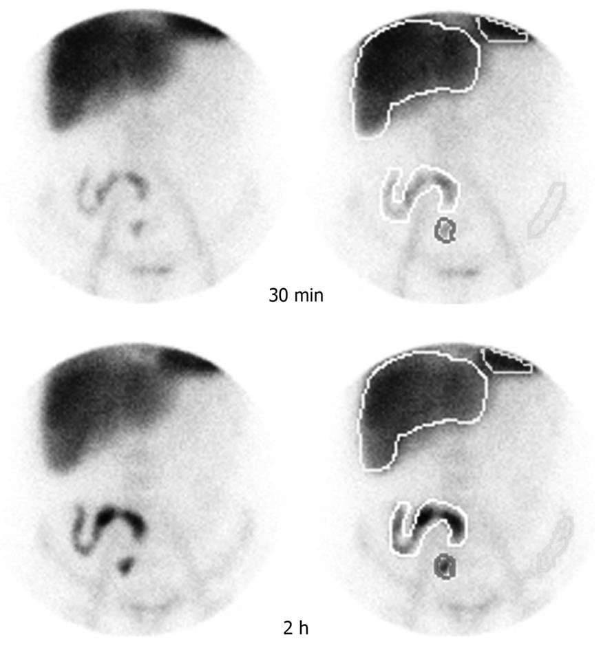
SI was calculated according to the method of Ybern et al[18]. Briefly, regions of interest (ROIs) were outlined over the liver, spleen, iliac crest and abnormal accumulations when present (Figure 1). The processing program of the gamma camera furnished the number of counts/area proportional to the radioactivity and the average value of the activity per pixel present in each region. The abdomen was divided into five zones: right, top, left, bottom and center, which corresponded approximately to the ascending colon, transverse colon, descending colon, sigmoid colon/rectum and small bowel. The SI was calculated in all scans in the anterior view: SI = (ΣAi) + B. “Ai” represents the degree of activity of the accumulations in each zone (1 = activity less than bone activity; 2 = activity greater than bone activity; 3 = activity greater than liver activity; 4 = activity greater than spleen activity). “B” indicates the number of zone with abnormal accumulations of labeled leukocytes (1 = one or more accumulations in one zone; 2 = accumulations in two or three zones; 3 = abnormal accumulations in four or five zones). SI > 2 was considered as active disease[18].
CDAI, SI and CRP were compared using the Spearman’s rank correlation.
The radiochemical purity of the lipophilic complex 99mTc-HMPAO presented a mean labeling percentage of the order of 85.0% ± 9.0% for the 25 samples. The mean yield for labeling of the autologous leukocytes with the lipophilic complex 99mTc-HMPAO was 55.0% ± 10.0%.
Six of the 20 patients (30%) presented congruent results for the three parameters investigated (CDAI, SI and CRP), which were two patients (I.M.F.; G.E.S.F.) with inflammatory activity and four (J.A.N.; F.R.F.; R.M.M.; A.B.O.) with disease in remission. On the other hand, 14 patients (70%) did not show congruent results for CDAI, SI and CRP (Table 1). Twelve patients showed results that were congruent with the Vienna Classification (Table 1) and technetium-99m-HMPAO-labeled leukocyte scintigraphy with regard to disease location. Besides, there was no correlation for both parameters that were described above in five patients; since scintigraphy images showed positive areas while these regions were not included in the Vienna Classification. In three other patients, the scintigraphy results did not show radioactivity uptake in the intestinal segments.
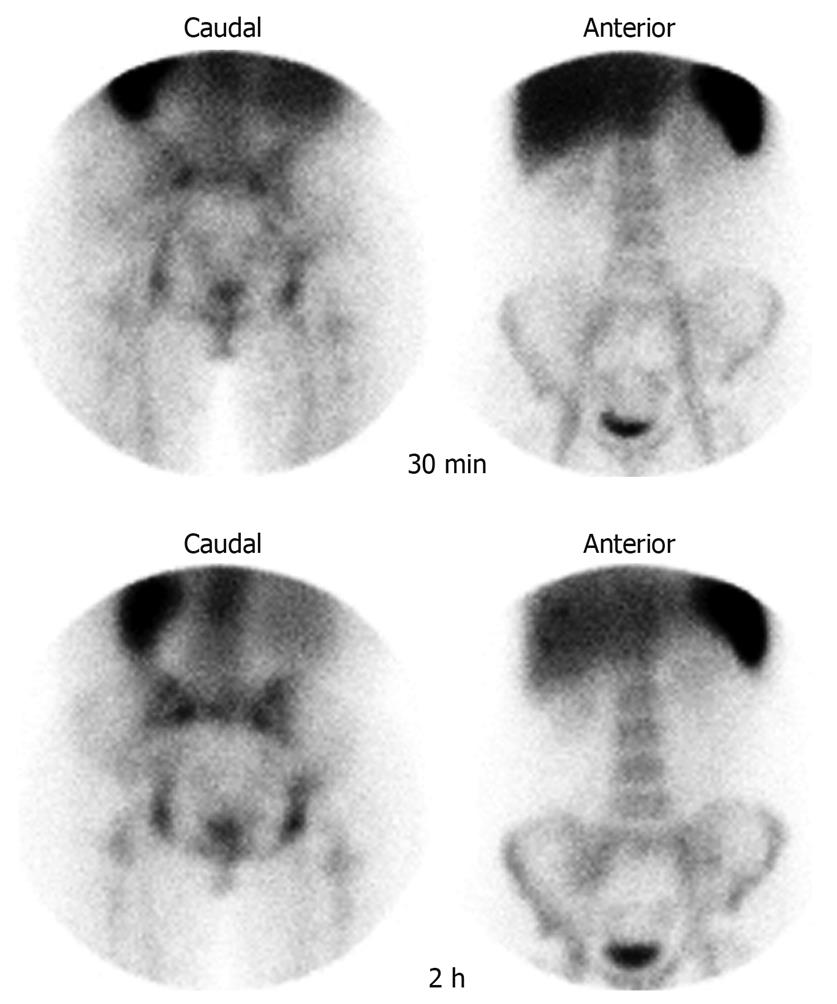
The images from a healthy volunteer in the anterior and caudal projections at 30 min and 2 h after injection of 99mTc-HMPAO-labeled leukocytes are presented in Figure 2. Intense accumulation in the liver and the spleen could be seen at 30 min post-injection, as well as accumulation in the bone marrow. Because it was a control case, there was no abnormal accumulation in the digestive tract.
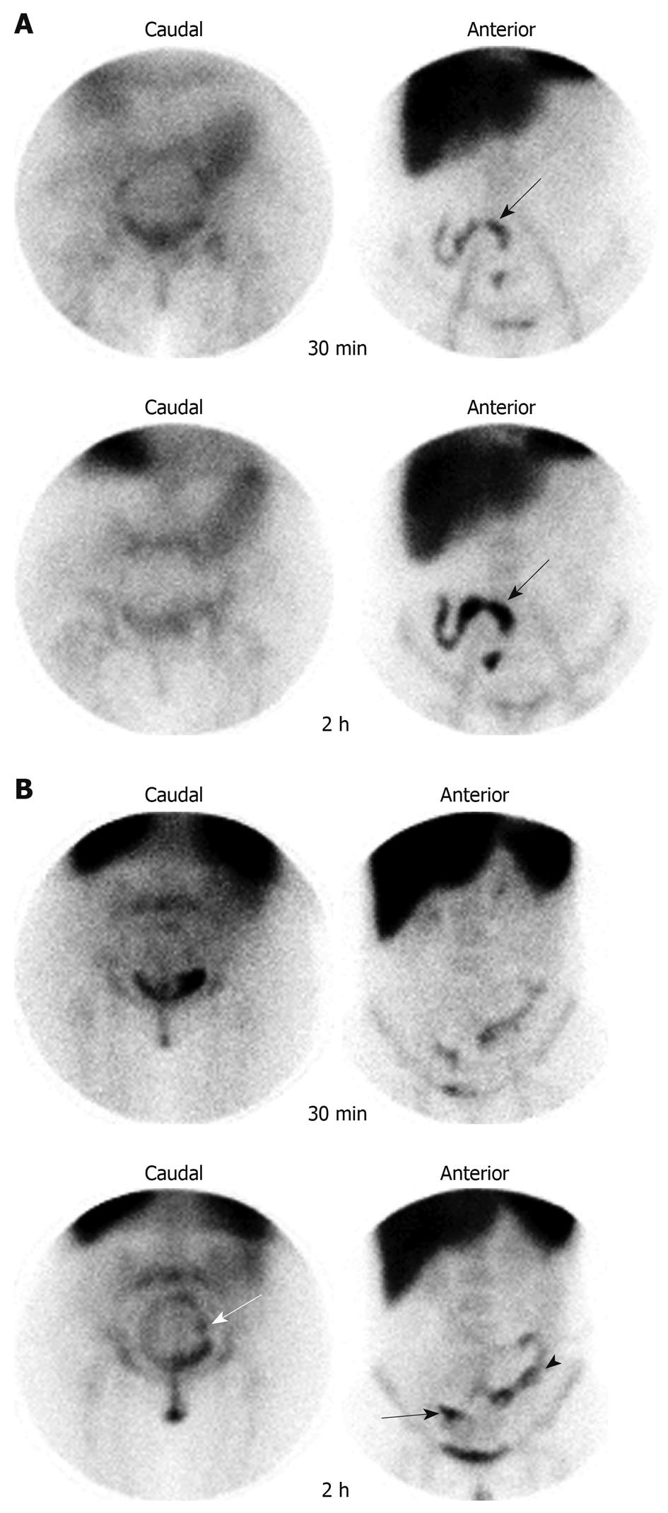
Accumulation of 99mTc-HMPAO-labeled leukocytes in the region of the terminal ileum, which suggested the presence of an inflammatory process at this location (anterior view), is shown in Figure 3A. At 2 h, uptake of labeled leukocytes increased, which showed the concentration of radiotracer in the indicated region. No pathological accumulation of 99mTc-HMPAO-labeled leukocytes could be seen in the caudal view, which indicated that there was no inflammation in the sigmoid and/or rectum.
The presence of inflammatory foci in the terminal ileum, descending colon, sigmoid and rectum, revealed by the intense accumulation of radiolabeled leukocytes, is shown in Figure 3B. Regions of the sigmoid and rectum affected by inflammation can be seen in the caudal projection. An increase in the radioactivity with time can also be seen.
According to the Spearman test, there was no significant correlation between the CDAI, SI and CRP in any of the investigated cases (Table 2).
| Spearman’s rank | Correlation coefficient (ρ) | ||
| t | P | ||
| CDAI and SI | 0.1835 | 0.7918 | 0.4388 |
| CDAI and CRP | 0.3373 | 1.5204 | 0.1457 |
| SI and CRP | 0.2783 | 1.2293 | 0.2347 |
The radiochemical purity of the lipophilic complex (HMPAO) labeled with technetium-99m was 85.0% ± 9.0%. The data suggest that 85% of the technetium-99m atoms were bound to HMPAO molecules. This result is in agreement with other data described in the literature[15]. On the other hand, the labeling yield for leukocytes was 55.0% ± 10%. Labeling yields of 46% and 65.5% have been reported previously[19,20]. Thus, the value obtained in the present work is supported by published data, which suggests that the manipulation process utilized in the preparation of the radiolabeled cells was adequate.
99mTc-HMPAO granulocyte scintigraphy of a healthy volunteer (Figure 2) showed uptake of radiolabeled leukocytes by the liver, spleen and bone marrow, which reflected physiological retention of labeled white blood cells[11].
The scintigraphic images were based on physiological alterations such as an increase in blood flow, vasodilatation, increased permeability and cellular leakage, which permit the precocious detection of inflammatory foci. These findings support the use of 99mTc-HMPAO granulocyte scintigraphy as an important examination for diagnostic screening of inflammatory diseases[4]. Cardoso et al[19] have observed sensitivity, specificity and accuracy values of > 87.5% in studies performed in patients highly suspected of having inflammatory bowel disease.
The data obtained in the present study showed that, in 70% of investigated cases, the results did not show correspondence with CDAI, CRP and SI (Table 1). It is known that CDAI is quite subjective and is based on a group of signs and clinical symptoms associated with erythrocyte sedimentation rate. This method was able to indicate inflammatory activity in four patients, while the SI and CRP indicated inflammatory activity in 13 and nine patients, respectively. Therefore, it is reasonable to suppose that the intestines of patients may have inflammatory foci that attract radiolabeled leukocytes in the specific case of CD, which results in a positive SI, although the signs and symptoms may still be absent.
As example of this observation, the patient L.M.T. (female, 54 years, Figure 3A) showed retention of radiolabeled leukocytes in the terminal ileum. On the other hand, the CDAI index (59.3) indicated disease remission and the CRP value (7.3 mg/L) suggested the absence of inflammatory activity. This patient was receiving treatment with prednisone and azathioprine, but inflammatory activity was present, which justified the uptake of 99mTc-HMPAO-labeled leukocytes in the region, however, the patient was asymptomatic.
A case that deserves special attention is the pathological accumulation of radiolabeled leukocytes in the terminal ileum, descending colon, sigmoid and rectum, which resulted in an SI of 8 (disease activity) as illustrated in Figure 3B (patient L.D., female, 24 years). Before performing 99mTc-HMPAO-labeled leukocyte scintigraphy, it was known that the CD in this patient was located only in the region of the terminal ileum, with absence of clinical signs, and the patient was receiving immunomodulator treatment. Seven days before 99mTc-HMPAO granulocyte scintigraphy, CRP level was elevated (34.1 mg/L). Despite all the segments that were identified by scintigraphy, the disease was considered to be in remission according to the CDAI (47.1). Thus, this result showed the importance of 99mTc-HMPAO-labeled leukocyte scintigraphy as a early diagnosis method for identifying affected regions not previously known. In addition, the therapeutic response should be also evaluated using this method, since the dose of immunomodulator administered to the patient probably was not sufficient to control the disease[11,12].
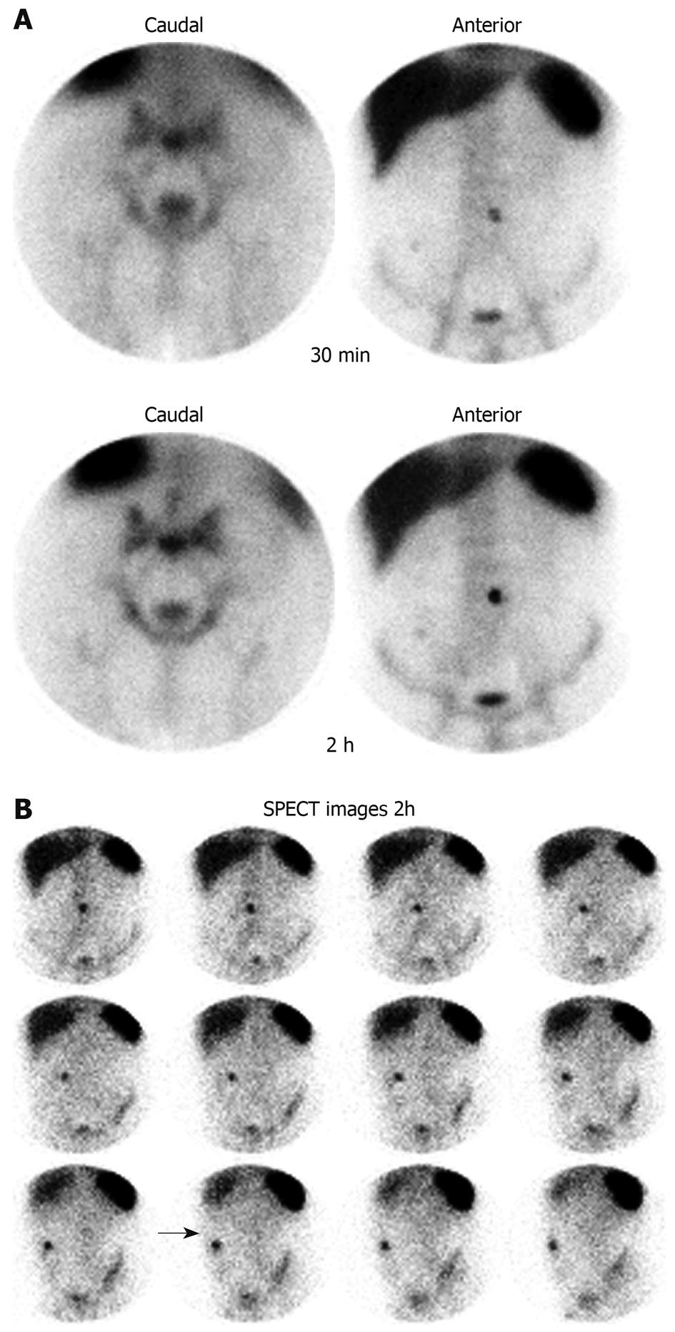
99mTc-HMPAO-labeled leukocyte scintigraphy performed in the patient J.A.N. (male, 44 years; Figure 4A) showed uptake of radiolabeled leukocytes in the central region of the abdomen, which indicated the presence of enterocutaneous fistula. On the other hand, the results did not show uptake of 99mTc-HMPAO-labeled leukocytes in segments of the intestine. SPECT showed that this radioactive uptake in the planar images corresponded to the external surface of the patient’s enterocutaneous fistula (Figure 4B). According to Arndt et al[10], the capture of leukocytes in areas outside the intestinal segments, such as abscesses and fistulas, should be analyzed separately, but not considered for the calculation of SI[10]. In our opinion, this uptake should be considered for SI calculation, because a fistula is a clinical feature that is suggestive of inflammatory activity and possible complications of CD.
The data obtained in the present work show that 99mTc-HMPAO-labeled leukocyte scintigraphy of the intestine could be useful for the evaluation of CD inflammatory activity, even in the absence of clinical signs and symptoms (disease remission). A functional method (scintigraphy) was compared with subjective (CDAI) and nonspecific (CRP) tests. Therefore, further studies will be necessary to prove the real utility of radiolabeled leukocytes to diagnosis and monitor patients with CD. Thus, it will be interesting to compare the scintigraphic method with another examination such as CT enterography.
technetium-99m-hexamethylpropyleneamine oxime (HMPAO)-labeled leukocyte scintigraphy was able to identify inflammatory activity in patients with Crohn’s disease (CD) even in the absence of signs and clinical symptoms.
Scintigraphic images are based on functional alterations of the tissues, and permit the early diagnosis of inflammatory bowel disease, when the anatomical alterations are not visible.
In the authors’ clinical routine, the Crohn’s disease activity index is used normally to evaluate the presence of inflammatory activity in patients. However, this index is subjective and sometimes is not able to detect the presence of inflammatory activity in subclinical disease. Thus, 99mTc-HMPAO granulocyte scintigraphy could contribute to follow-up of these patients.
The results obtained suggest that 99mTc-HMPAO-labeled leukocyte scintigraphy could be used to monitor inflammatory bowel disease and evaluate the efficiency of therapy.
Scintigraphic images are obtained using compounds or cells labeled with radioactive isotopes such as technetium-99m, iodine-131 and gallium-67. CD is a chronic intestinal inflammation of unknown etiology that can affect any segments of the digestive tract.
The number of patients is small and therefore their positive experience with the technique may decrease with time and number of patients. The figures are of high quality and indicate the expertise of the authors.
Peer reviewer: Amado S Peña, Professor, Department of Pathology, Immunogenetics, VU University Medical Centre, De Boelelaan 1117, PO Box 7057, Amsterdam 1007 MB, The Netherlands
S- Editor Wang YR L- Editor Kerr C E- Editor Lin YP
| 1. | Crawford JM, Kumar V. The oral cavity and the gastrointestinal tract. Basic Pathology. Philadelphia: WB Saunders 2003; 543-590. |
| 2. | Carter MJ, Lobo AJ, Travis SP. Guidelines for the management of inflammatory bowel disease in adults. Gut. 2004;53 Suppl 5:V1-V16. |
| 3. | Damião AOMC, Sipahi AM. Doença inflamatória intestinal. Gastroenterologia. Rio de Janeiro: Medsi Editora Médica e Científica Ltda 2004; 1105-1149. |
| 4. | Stathaki MI, Koukouraki SI, Karkavitsas NS, Koutroubakis IE. Role of scintigraphy in inflammatory bowel disease. World J Gastroenterol. 2009;15:2693-2700. |
| 5. | Stange EF, Travis SP, Vermeire S, Beglinger C, Kupcinkas L, Geboes K, Barakauskiene A, Villanacci V, Von Herbay A, Warren BF. European evidence based consensus on the diagnosis and management of Crohn's disease: definitions and diagnosis. Gut. 2006;55 Suppl 1:i1-i15. |
| 6. | Annovazzi A, Biancone L, Caviglia R, Chianelli M, Capriotti G, Mather SJ, Caprilli R, Pallone F, Scopinaro F, Signore A. 99mTc-interleukin-2 and (99m)Tc-HMPAO granulocyte scintigraphy in patients with inactive Crohn's disease. Eur J Nucl Med Mol Imaging. 2003;30:374-382. |
| 7. | Best WR, Becktel JM, Singleton JW, Kern F Jr. Development of a Crohn's disease activity index. National Cooperative Crohn's Disease Study. Gastroenterology. 1976;70:439-444. |
| 8. | Rennen HJ, Boerman OC, Oyen WJ, Corstens FH. Imaging infection/inflammation in the new millennium. Eur J Nucl Med. 2001;28:241-252. |
| 9. | Peters AM, Danpure HJ, Osman S, Hawker RJ, Henderson BL, Hodgson HJ, Kelly JD, Neirinckx RD, Lavender JP. Clinical experience with 99mTc-hexamethylpropylene-amineoxime for labelling leucocytes and imaging inflammation. Lancet. 1986;2:946-949. |
| 10. | Arndt JW, Grootscholten MI, van Hogezand RA, Griffioen G, Lamers CB, Pauwels EK. Inflammatory bowel disease activity assessment using technetium-99m-HMPAO leukocytes. Dig Dis Sci. 1997;42:387-393. |
| 11. | Martin-Comin J, Prats E. Clinical applications of radiolabeled blood elements in inflammatory bowel disease. Q J Nucl Med. 1999;43:74-82. |
| 12. | Capdevila CB. Enfermedad inflamatoria intestinal. Diagnóstico de la inflamación y de la infección en Medicina Nuclear. Madrid: SIMED Software S.L 2005; 25-51. |
| 13. | Martin-Comin J, Cardoso VN, Plaza P, Roca M. Hanks balanced salt solution: an alternative resuspension medium to label autologous leukocytes. Experience in inflammatory bowel disease. Braz Arch Biol Technol. 2002;45:39-44. |
| 14. | Roca M, Martín-Comín J, Becker W, Bernardo-Filho M, Gutfilen B, Moisan A, Peters M, Prats E, Rodrigues M, Sampson C. A consensus protocol for white blood cells labelling with technetium-99m hexamethylpropylene amine oxime. International Society of Radiolabeled Blood Elements (ISORBE). Eur J Nucl Med. 1998;25:797-799. |
| 15. | Barthel H, Kämpfer I, Seese A, Dannenberg C, Kluge R, Burchert W, Knapp WH. Improvement of brain SPECT by stabilization of Tc-99m-HMPAO with methylene blue or cobalt chloride. Nuklearmedizin. 1999;38:80-84. |
| 16. | Biancone L, Schillaci O, Capoccetti F, Bozzi RM, Fina D, Petruzziello C, Geremia A, Simonetti G, Pallone F. Technetium-99m-HMPAO labeled leukocyte single photon emission computerized tomography (SPECT) for assessing Crohn's disease extent and intestinal infiltration. Am J Gastroenterol. 2005;100:344-354. |
| 17. | Martin-Comin J, Brulles YR, Salvadó JM, Lázaro MTB, Engronyat MR, Añé RP. La Medicina Nuclear en el diagnóstico de la enfermedad inflamatoria intestinal. Diagnóstico de la inflamación y de la infección en Medicina Nuclear. Madrid: SIMED Software S.L 2005; 131-144. |
| 18. | Ybern A, Martin-Comin J, Giné JJ, Casanovas T, Villa R, Gassull MA. 111In-oxine-labelled autologous leucocytes in inflammatory bowel disease: new scintigraphic activity index. Eur J Nucl Med. 1986;11:341-344. |
| 19. | Cardoso VN, Plaza PJ, Roca M, Armero F, Martín-Comin J. Assessment of inflammatory bowel disease by using two different (99m)Tc leucocyte labelling methods. Nucl Med Commun. 2002;23:715-720. |
| 20. | Arndt JW, van der Sluys Veer A, Blok D, Griffoen G, Verspaget HW, Lamers CB, Pauwels EK. Prospective comparative study of technetium-99m-WBCs and indium-111-granulocytes for the examination of patients with inflammatory bowel disease. J Nucl Med. 1993;34:1052-1057. |


