Published online Jan 21, 2009. doi: 10.3748/wjg.15.378
Revised: December 10, 2008
Accepted: December 17, 2008
Published online: January 21, 2009
Although Bouveret’s syndrome, i.e. gastric outlet obstruction by a large gallstone impacted in the proximal duodenum secondary to a cholecystoduodenal fistula, is rare, its pathogenesis and clinical features are well characterized. However, existence of variant forms of the syndrome are not well known, and as far as we know, only two cases of variant Bouveret’s syndrome have been described in the English-language literature. We present a case of another new variant of Bouveret’s syndrome in a 54-year-old Korean woman.
- Citation: Park SH, Lee SW, Song TJ. Another new variant of Bouveret’s syndrome. World J Gastroenterol 2009; 15(3): 378-379
- URL: https://www.wjgnet.com/1007-9327/full/v15/i3/378.htm
- DOI: https://dx.doi.org/10.3748/wjg.15.378
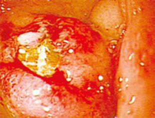
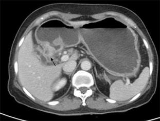
We read with much interest the case report of Arioli et al[1], ‘a new variant of Bouveret’s syndrome’, recently published in World Journal of Gastroenterology. They described a case of gallstone ileus in which an incomplete pyloric occlusion developed following the migration of a large stone through a cholecystogastric fistula, in a patient with undiagnosed chronic cholecystitis. In their case, the gallstone lay in the gastric antrum rather than within the lumen of the duodenum, which is different from typical Bouveret’s syndrome. Doody et al[2] have also described a variant of Bouveret’s syndrome in which duodenal obstruction was caused by a huge gallstone that lay outside the duodenum, which gave an atypical Bouveret’s syndrome appearance. We recently encountered a similarly interesting case of gastric outlet obstruction caused by complicated gallstone disease. A 54-year-old Korean woman was admitted to the hospital with epigastric pain and intermittent vomiting of 20 d duration. She had a history of recurrent bouts of right upper quadrant abdominal pain caused by acute cholecystitis. Gastroduodenoscopy showed an obstructive duodenal mass, the center of which harbored a mucosal defect (Figure 1). Abdominal computed tomography (CT) showed a cystic lesion within the thickened wall of the bulbar and post-bulbar portions of the duodenum (Figure 2). The gallbladder contained a small stone and was partially collapsed. On laparotomy, the gallbladder was densely adhered to the first portion of the duodenum. The patient underwent cholecystectomy, with excision of a portion of the duodenal wall. In the cystic lesion of the duodenal wall, muddy stone fragments were found. Based on operative findings, it was certain that a large gallstone had passed through the cholecystoduodenal fistula into the duodenal wall, and remained there sufficiently long to form a submucosal mass that eroded through the mucosa into the duodenal lumen. Although the stone was passed without causing any further problems, the cavaitary lesion in the duodenal wall persisted and bulged into the duodenal lumen, which caused the duodenal obstruction. The features of our patient resembled a Bouveret’s syndrome in clinical presentation, but differed from it inasmuch that the duodenal obstruction was not caused by a gallstone per se, but rather by secondary changes in the duodenal wall caused by a gallstone. A cystic lesion within the wall of the duodenum caused confusion in our case, and surgical exploration was required to make a definite diagnosis and to determine a treatment strategy. We think that this case may be another new variant of Bouveret’s syndrome.
| 1. | Arioli D, Venturini I, Masetti M, Romagnoli E, Scarcelli A, Ballesini P, Borghi A, Barberini A, Spina V, De Santis M. Intermittent gastric outlet obstruction due to a gallstone migrated through a cholecysto-gastric fistula: a new variant of "Bouveret's syndrome". World J Gastroenterol. 2008;14:125-128. |


