Published online Aug 14, 2008. doi: 10.3748/wjg.14.4838
Revised: June 30, 2008
Accepted: July 7, 2008
Published online: August 14, 2008
The patient was a 33-year-old man with hematochezia. Colonoscopy revealed a lobulated peduncular polyp with bleeding, about 40 mm in diameter, in the ascending colon. The polyp had both red and white components and a mosaic pattern. Magnifying observation revealed a red rugged surface component, and smooth white nodules with enlarged round or oval crypt openings. Endoscopic polypectomy was performed. Histological examination of the specimen revealed inflammatory granulation tissue in the lamina propria, proliferation of smooth muscle, and hyperplastic glands with cystic change. This polyp was diagnosed as inflammatory myoglandular polyp (IMGP). Lobulated-type IMGP in the ascending colon is rare.
- Citation: Kanzaki H, Hirasaki S, Okuda M, Kudo K, Suzuki S. Lobulated inflammatory myoglandular polyp in the ascending colon observed by magnifying endoscopy and treated with endoscopic polypectomy. World J Gastroenterol 2008; 14(30): 4838-4840
- URL: https://www.wjgnet.com/1007-9327/full/v14/i30/4838.htm
- DOI: https://dx.doi.org/10.3748/wjg.14.4838
Inflammatory myoglandular polyp (IMGP) is characterized by inflammatory granulation tissue in the lamina propria[1], proliferation of smooth muscle[2], and hyperplastic glands with variable cystic change[3-7]. Only a small number of cases have been reported, and its pathogenesis and natural history remain unclear[8-12]. Herein, we describe a relatively rare case of loburated-type IMGP in the ascending colon causing hematochezia. We also report the magnifying endoscopy findings of this IMGP.
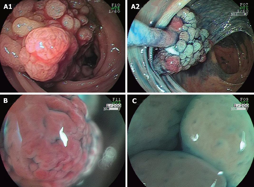
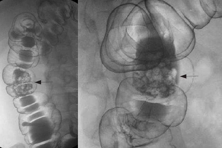
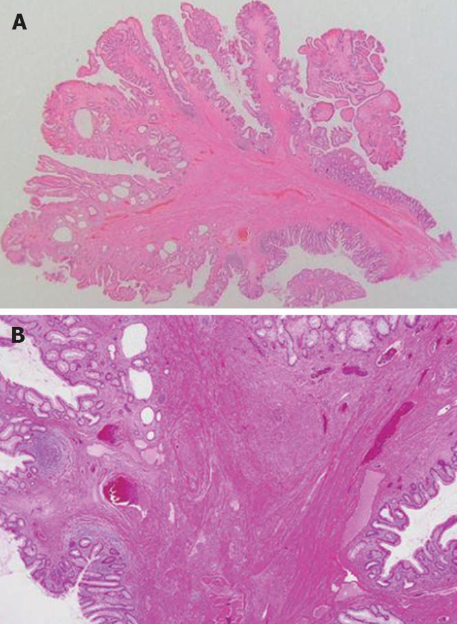
A 33-year-old man presented with the symptom of hematochezia. He was diagnosed with chronic renal failure and underwent hemodialysis 5 years ago. His body temperature was 36.7°C, blood pressure was 148/82 mmHg, and radial pulse rate was 70 beats/min and regular. He had anemia. Laboratory tests showed hemoglobin concentration of 8.0 g/dL [normal range (NR): 12-16 g/dL], a red blood cell count of 325 × 104 /μL (NR: 380-500 × 104 /μL), a white blood cell count of 8400/μL, and a platelet count of 28.8 × 10⁴/μL. The levels of hepatic and biliary enzymes, such as aspartate aminotransferase (AST), alanine aminotransferase (ALT), alkaline phosphatase (ALP), γ-glutamyltranspeptidase (γ-GTP), and lactate dehydrogenase (LDH), were normal. On renal function tests, the blood urea nitrogen and creatinine levels were 44.9 mg/dL (NR: 8-20 mg/dL) and 14.1 (NR: 0.5-1.3 mg/dL), respectively. Colonoscopy revealed a lobulated peduncular polyp with bleeding, about 40 mm in diameter, in the ascending colon (Figure 1A1). The polyp had both red and white components and a mosaic pattern (Figure 1A2). Magnifying observation (EC-450ZH, Fujinon Toshiba ES Systems) revealed a red, slightly rugged surface component without normal mucosal structure (Figure 1B), and smooth white nodules with enlarged round or oval crypt openings (Figure 1C). We speculated that this polyp was non-neoplastic. It was suspected to be an inflammatory polyp from endoscopic findings. An air contrast barium enema also revealed a pedunculated, lobulated polyp in the ascending colon (Figure 2). Endoscopic polypectomy was performed. Histological examination of the specimen revealed inflammatory granulation tissue in the lamina propria, proliferation of smooth muscle, and hyperplastic glands with cystic change (Figure 3). The lesion was diagnosed as IMGP. After endoscopic polypectomy, the symptom of hematochezia was resolved.
IMGP is a non-neoplastic colorectal polyp, first described by Nakamura et al[1]. IMGP is solitary, pedunculated and rarely covered by a fibrin cap, and follows a benign course. Also, IMGP has no association with inflammatory bowel diseases and is located not only in the rectosigmoid, but also in the descending and transverse colon[3].
Only a small number of IMGP cases have been reported. According to Fujino et al[8], a review of the literature revealed 48 cases of IMGP in the large intestine up to 2001. However, recent advances in diagnostic techniques have enabled us to identify small and asymptomatic polyps, and reports on IMGP of the colon have been increasing[9,10]. Fujino et al[8] described that the macroscopic appearance was the pedunculated type in 83.3% of cases. In that report, the sites of IMGP in the large intestine were studied and 47 of 48 cases (97.9%) had lesions in the rectum to transverse colon. Thus, IMGPs of the large intestine are predominantly in the distal colon[8,9].
IMGPs in the colon are usually asymptomatic and often detected incidentally on barium enema or endoscopy[8-10]. Another review of the literature revealed that the main clinical feature of colorectal IMGPs is hematochezia[10,11]. Endoscopic characteristic findings are as follows: (1) pedunculated or semipedunculated, (2) red and (3) smooth, spherical and hyperemic surface with patchy mucous exudation and erosion[8,9]. IMGPs should be distinguished from other colorectal polyps such as inflammatory fibroid polyps (IFP)[10,13], Peutz-Jegher-type polyps or juvenile polyps[10,11]. However, the correct endoscopic diagnosis of colorectal IMGP can seldom be made. Endoscopic findings of IMGP are similar to those of IFP and juvenile polyp. The final diagnosis of colonic IMGP depends on the pathological findings of endoscopic mucosal resection (EMR) or endoscopic polypectomy specimens. Harada et al[7] reviewed the literature and described that 4 of 40 IMGPs (10%) were lobulated. The present case was rare because IMGP located in the ascending colon and the radiographic and endoscopic findings of the polyp were the lobulated type.
Moriyama et al[9] reported magnifying endoscopic findings of 5 IMGPs in 2003. They described that magnifying observation revealed a slightly rugged surface consisting of aggregated smooth nodules with enlarged round or oval crypt openings. In the present case, magnifying endoscopic findings were the same as Moriyama’s description. However, this polyp was unique because the lesion had red and white components and a mosaic pattern. There have been few reports on magnifying endoscopic findings of IMGP. To clarify the characteristic magnifying endoscopic findings of IMGP, we should accumulate and analyze many cases of IMGP.
As to therapy, IMGP of the large intestine can best be removed endoscopically, because it is thought to be clinically and histologically benign. Most Japanese cases are treated with polypectomy or EMR[5,7-10]. Endoscopic or surgical treatment is necessary if gastrointestinal bleeding[9] or colonic intussusception occurs. Local excision of the polyp is curative. Kayhan et al[12] reported a case of large IMGP (> 6 cm in diameter), which was too large for endoscopic removal, and treated with surgical resection. We consider that the percentage of colonic IMG patients who undergo surgical resection will decrease and endoscopic resection will increase in the future because of advances in diagnostic techniques such as improved endoscopic images and the discovery of asymptomatic small IMGPs.
In conclusion, we reported a case of IMGP in the ascending colon causing hematochezia. IMGP should generally be taken into consideration as a differential diagnosis of peduncular polyp of the colon. IMGP of the large intestine is not fatal and patients remain asymptomatic in their daily lives except for gastrointestinal bleeding or bowel obstruction. Therefore, it is likely that there will be many latent patients with IMGP in the future. Endoscopists should be aware of IMGP endoscopic characteristics, although lobulated type-IMGP in the ascending colon is rare. Further studies on the magnifying endoscopy findings of IMGP are certainly required.
Peer reviewer: William Dickey, Professor, Altnagelvin Hospital, Londonderry, Northern Ireland BT476SB, United Kingdom
S- Editor Li DL L- Editor Wang XL E- Editor Yin DH
| 1. | Nakamura S, Kino I, Akagi T. Inflammatory myoglandular polyps of the colon and rectum. A clinicopathological study of 32 pedunculated polyps, distinct from other types of polyps. Am J Surg Pathol. 1992;16:772-779. |
| 2. | Griffiths AP, Hopkinson JM, Dixon MF. Inflammatory myoglandular polyp causing ileo-ileal intussusception. Histopathology. 1993;23:596-598. |
| 3. | Bhathal PS, Chetty R, Slavin JL. Myoglandular polyps. Am J Surg Pathol. 1993;17:852-853. |
| 4. | Gómez Navarro E, del Río Martín JV, Sarasa Corral JL, Melero Calleja E. [Myoglandular inflammatory polyp located in the distal end of the rectum]. Rev Esp Enferm Dig. 1994;85:45-46. |
| 5. | Nagata S, Sumioka M, Sato O, Miyamoto M, Watanabe C, Yamada H, Hirata K, Imagawa M, Haruma K, Kajiyama G. [Five cases of inflammatory myoglandular polyp]. Nippon Shokakibyo Gakkai Zasshi. 1998;95:145-150. |
| 6. | Bhardwaj K, Mohan H, Chopra R, Bhardwaj S, Sachdev A. Inflammatory myoglandular polyp of rectum. Indian J Gastroenterol. 1998;17:63-64. |
| 7. | Harada N, Chijiiwa Y, Yao T, Koyanagi M, Ono Y, Motomura S. Inflammatory myoglandular polyp. J Clin Gastroenterol. 1999;29:104-105. |
| 8. | Fujino Y, Orii S, Nakamura S, Sugai T, Saito S, Yamaguchi T, Noro S, Ishii M, Inomata M, Suzuki K. Five cases of colorectal inflammatory myoglandular polyps. Gastroenterological Endoscopy. 2001;43:1281-1286. |
| 9. | Moriyama T, Matsumoto T, Hizawa K, Tada S, Fuchigami T, Iwai K, Yao T, Iida M. Inflammatory myoglandular colorectal polyps: a case series of nine patients. Endoscopy. 2003;35:363-365. |
| 10. | Tashiro M, Yoshikawa I, Matsuhashi T, Yamasaki T, Nishikawa S, Taguchi M, Yamasaki M, Kume K, Otsuki M. Images of interest. Gastrointestinal: inflammatory myoglandular polyp of the colon. J Gastroenterol Hepatol. 2005;20:1123. |
| 11. | Becheanu G, Stamm B. Inflammatory myoglandular polyp--a rare but distinct type of colorectal polyps. Pathol Res Pract. 2003;199:837-839. |
| 12. | Kayhan B, Küçükel F, Akdoğan M, Ozaslan E, Küçükbaş TA, Atoğlu O. Inflammatory myoglandular polyp: a rare cause of hematochezia. Turk J Gastroenterol. 2004;15:117-119. |
| 13. | Hirasaki S, Matsubara M, Ikeda F, Taniguchi H, Suzuki S. Inflammatory fibroid polyp occurring in the transverse colon diagnosed by endoscopic biopsy. World J Gastroenterol. 2007;13:3765-3766. |


