Published online May 14, 2008. doi: 10.3748/wjg.14.2877
Revised: February 4, 2008
Published online: May 14, 2008
AIM: To investigate the influence of different quasispecies of hepatitis C virus (HCV) genotype 1b core protein on growth of Chang liver cells.
METHODS: Three eukaryotic expression plasmids (pEGFP-N1/core) that contained different quasispecies truncated core proteins of HCV genotype 1b were constructed. These were derived from tumor (T) and non-tumor (NT) tissues of a patient infected with HCV and C191 (HCV-J6). The core protein expression plasmids were transiently transfected into Chang liver cells. At different times, the cell cycle and apoptosis was assayed by flow cytometry, and cell proliferation was assayed by methyl thiazolyl tetrazolium (MTT) assay.
RESULTS: The proportion of S-phase Chang liver cells transfected with pEGFP-N1/core was significantly lower than that of cells transfected with blank plasmid at three different times after transfection (all P < 0.05). The proliferation ratio of cells transfected with pEGFP-N1/core was significantly lower than that of cells transfected with blank plasmid. Among three different quasispecies, T, NT and C191 core expression cells, there was no significant difference in the proportion of S- and G0/G1-phase cells. The percentage of apoptotic cells was highest for T (T > NT > C191), and apoptosis was increased in cells transfected with pEGFP-N1/core as the transfection time increased (72 h > 48 h > 24 h).
CONCLUSION: These results suggest that HCV genotype 1b core protein induces apoptosis, and inhibits cell-cycle progression and proliferation of Chang liver cells. Different quasispecies core proteins of HCV genotype 1b might have some differences in the pathogenesis of HCV persistent infection and hepatocellular carcinoma.
- Citation: Yan XB, Mei L, Feng X, Wan MR, Chen Z, Pavio N, Brechot C. Hepatitis C virus core proteins derived from different quasispecies of genotype 1b inhibit the growth of Chang liver cells. World J Gastroenterol 2008; 14(18): 2877-2881
- URL: https://www.wjgnet.com/1007-9327/full/v14/i18/2877.htm
- DOI: https://dx.doi.org/10.3748/wjg.14.2877
Hepatitis C virus (HCV) infection is prevalent worldwide. HCV infection may lead to the development of chronic hepatitis, cirrhosis and hepatocellular carcinoma (HCC)[1]. HCV is a positive-strand RNA virus that belongs to the family Flaviviridae. The HCV RNA genome is 9.6 kb in length and encodes a precursor polyprotein of 3000 amino acids. The precursor polyprotein is cleaved by host and viral proteases and produces a series of structural and non-structural proteins. In addition, HCV genome has the property of variation, and it usually exists as different quasispecies of the same genotype in HCV-infected patients[23]. The function of different quasispecies of HCV is not fully identical[45].
HCV core protein is important as a viral structural component in nucleocapsid formation, and it is highly conserved among different HCV genotypes. Several studies have established that HCV core protein can regulate cell signal transduction, such as MAPK-, JAK-STAT-, NF-κB-, AP-1- and SRE-associated pathways[6–8]. Hence, it plays important roles in the pathogenesis of HCV persistent infection, as well as HCC. However, the mechanism of persistent infection and HCC due to HCV core protein infection remains unclear. Although several studies have investigated the influence of the HCV core protein on cell growth and apoptosis, the results of these investigations have been inconsistent and mutually conflicting.
In most studies, HCV core protein has been expressed in single clones of permanently transfected cell lines, while more recently, transiently transfected cells have also been used. These studies have found that core protein affects normal cellular functions, such as proliferation and death, which are involved, directly or indirectly, in HCV hepatocarcinogenesis. Because we have found that different quasispecies core proteins of HCV genotype 1b have some differences in the pathogenesis of HCV persistent infection and HCC. HCV is a hepatotropic virus, it is therefore more appropriate to use a hepatocyte cell line to study the function of HCV core protein.
To further clarify the in vivo role of truncated HCV core protein from different genotype 1b quasispecies, we expressed three different truncated forms of HCV core protein derived from tumor tissue (T), non-tumor tissue (NT) and C191 (HCV-J6), which all belong to HCV genotype 1b, in transiently transfected Chang liver cells, an immortalized non-tumor hepatic cell line. Cell cycle and apoptosis were assayed by flow cytometry, and cell proliferation was assayed by methyl thiazolyl tetrazolium (MTT) assay.
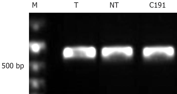
Three different truncated HCV core protein eukaryotic expression plasmids, pEGFP-N1/core, were constructed. Truncated core protein nucleotide sequences were amplified from pGEX 4T-1/HCV-core, which contained core sequences from T, NT and C191, respectively. Sequence analysis revealed that T and NT were all HCV genotype 1b. The primers were designed according to the core protein gene sequence of T, NT and C191 (Table 1). PCR reaction system: 50 &mgr;L: water 40.75 &mgr;L; PCR reaction buffer (10 ×), 5 &mgr;L; template (45 ng), 1 &mgr;L; primer up 20 pmol/L, 1 &mgr;L; primer down 20 pmol/L, 1 &mgr;L; dNTP 20 mmol/L, 1 &mgr;L, Expand high enzyme 0.25 &mgr;L, 94°C for 2 min, 94°C for 30 s, 50°C for 30 s, 72°C for 30 s, 10 cycles; 94°C for 30 s, 55°C for 30 s, 72°C for 30 s, 20 cycles; and 72°C for 7 min. PCR products (Figure 1) were purified and cleaved with restriction enzymes NheIand EcoRI, and cloned into pEGFP-N1 to yield GFP-core fusion protein plasmids.
| Primer up (location) | Primer down (location) | |
| T: 1-172 | CGCGCTAGCATGAGGCACGAATCC (1-14) | CCGGAATTCGCAACCGGGCAG (505-516) |
| NT: 1-172 | CGCGCTAGCATGAGCACGAATCC (1-14) | CCGGAATTCGGCAACCGGGCAGATTC (505-516) |
| C191: 1-172 | CGCGCTAGCATGAGCACAAATCC (1-14) | CCGGAATTCGGCAACCGGGCAAATTC (505-516) |
Chang liver cells were grown in Dulbecco’s modified Eagle’s medium (Gibco, USA) supplemented with 10% fetal bovine serum (FBS; Gibco) at 37°C in 5% CO2. Cells were incubated for 18 h and then transfected with Lipofectamine 2000 (Invitrogen, USA) following the manufacturer’s instructions. Two micrograms of plasmids were used to achieve transfection. After 6 h, the medium was changed and cells were collected at different times.
The cell cycle and apoptosis was assayed by flow cytometry at 24, 48 and 72 h after transfection. Ten × 105 cells were trypsinized, pelleted by centrifugation, and resuspended in PBS, and then cells were fixed with 70% ethanol and stored at 4°C overnight. After washing twice, cells were incubated in propidium iodine for 20 min at room temperature. The cell suspension was analyzed by flow cytometry using Cell Quest software (Becton Dickinson, USA).
Cell proliferation was assayed by MTT assay and a growth curve was created. 104 cells were seeded per well on a 96-well plate. Zero, 24, 48, 72 and 96 h after transfection, 10 &mgr;L water-soluble tetrazolium reagent (MTT; Sigma, USA) was added to 100 &mgr;L culture medium. Cells were then incubated at 37°C in 5% CO2 for 4 h. A570 was measured. The assay was done in quadruplicate.
The values were expressed as mean ± SD. Experimental data were analyzed by SPSS 13.0 software (SPSS Inc, Chicago, IL, USA). Comparison of different groups was analyzed by one-way analysis of variance (ANOVA). P < 0.05 was considered statistically significant.
As shown in Tables 2, 3, 4, three different quasispecies truncated core proteins inhibited Chang liver cell cycle progression by impairing G1- to S-phase transition. The proportion of S- and G0/G1-phase Chang liver cells transfected with pEGFP-N1/core was significantly lower than that of cells transfected with blank plasmid at 24, 48 and 72 h after transfection (P = 0.002, P = 0.001, P = 0.001, respectively), but there were no significant differences among cells expressing the three different quasispecies HCV truncated core proteins.
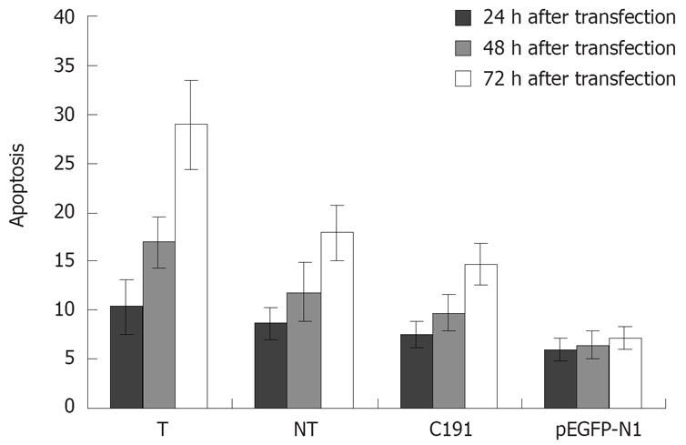
As shown in Figure 2, three different quasispecies truncated core proteins induced apoptosis at different levels. The apoptotic ratio of Chang liver cells transfected with pEGFP-N1/core was significantly higher than that of cells transfected with blank plasmid. The apoptotic percentage of T was the highest, and C191 was the lowest (T > NT > C191). The apoptosis ratio increased in cells transfected with pEGFP-N1/core as transfection time increased (72 h > 48 h > 24 h).
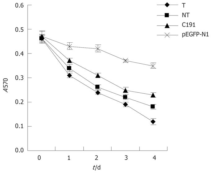
We found that different HCV genotype 1b quasispecies core proteins inhibited the Chang liver cell cycle by impairing G1- to S-phase transition and induced aptoptosis at different times after transfection. Chang liver cell proliferation was further analyzed using MTT assay. As shown in Figure 3, different HCV genotype 1b quasispecies core proteins inhibited Chang liver cell proliferation. Among the three different HCV genotype 1b quasispecies core proteins, that of T inhibited Chang liver cell proliferation more obviously than NT and C191 (T > NT > C191) at 24, 48, 72 and 96 h.
HCV core protein is a multifunctional protein that can modulate a number of cellular processes, including transcription, inhibition or stimulation of cell cycle and apoptosis, and suppression of host immunity. In this study, using flow cytometry and MTT assay, we investigated the influence of different quasispecies of truncated HCV genotype 1b core proteins on growth of Chang liver cells, an immortalized non-tumor hepatic cell line. We found that HCV genotype 1b core protein HCV induced apoptosis, and inhibited the cell cycle and cell proliferation. Among the three different quasispecies core proteins, the rates of inducing apoptosis and inhibiting cell proliferation were different, which suggests that different quasispecies of HCV genotype 1b core proteins might have some differences in their pathogenesis of HCV persistent infection and HCC.
Regulation of cell cycle and apoptosis is essential to maintain normal cell growth. Several studies have reported that HCV core protein can influence cell cycle progression. Two important checkpoints of the cell cycle, G1/S and G2/M, are responsible for accumulation of detrimental mutations that may result in cell transformation. The mechanism of cell cycle regulation is quite complex, and involves a number of proteins, such as p53, p21, PRb, E2F and c-myc[910]. Ruggieri et al have reported that stable expression of core protein in unsynchronized HepG2 cells induces perturbation of the cell cycle by increasing the S-phase fraction. c-myc protein stability is increased, which is one of the regulatory molecules of the cell cycle[11]. Honda et al have found that core protein promotes cell cycle progression in stable CHO transformation by upregulation of c-myc[12]. Ohkawa et al have reported that core protein impairs G1 to S transition by using core expression stable CL2 cell, a murine normal liver-derived cell line. E2F-mediated transcription, and PRb, CDK4 and CDK2 activities were suppressed by HCV core protein[13]. Different studies have come to different conclusions. We found that transient expression of HCV genotype 1b truncated core proteins could impair G1/S in the Chang liver cell cycle. Different quasispecies core proteins from T, NT and C191 have no significant difference when it comes to impairing G1/S. The above studies suggest that core protein might be a double-function protein, promoting cell cycle progression and inhibiting cell cycle progression. However, the exact mechanisms of how different quasispecies core proteins affect the G1/S checkpoint remain unclear.
Apoptosis is defined as programmed cell death and it is an orderly process. Apoptosis can be induced by a variety of stimuli, including oxidative stress, heat shock, ionizing radiation, cytokines and virus infection. The apoptotic process appears to be a host defense mechanism against viral infections. HCV might lead to viral persistence by inhibiting hepatocyte apoptosis and inducing monocyte apoptosis[14]. Although diverse effects of the core protein on apoptosis have been reported, the underlying mechanisms are not fully understood[15]. Core protein exhibits both pro- and anti-apoptotic actions. Transient expression of the core protein can inhibit tumor necrosis factor (TNF)-α-mediated apoptosis in MCF7 cells, a cell line derived from breast carcinoma tissue[16]. In contrast, several studies have indicated that core protein can promote apoptosis induced by Fas or TNF-α in stable expression systems in HepG2, HeLa and Jurkat T cell lines[17–19]. In our study, we found that different quasispecies of HCV genotype 1b truncated core proteins could induce Chang liver cell apoptosis, and the ability to induce apoptosis was different (T > NT > C191). Apoptosis was increased depending on prolongation of transfection time (72 h > 48 h > 24 h). Moreover, the cell proliferation induced by HCV core protein was consistent with our finding that HCV core protein induced cell apoptosis. Nevertheless, the present results allow us to propose an alternative, and in fact contrasting mechanism for HCV core protein. Our observations imply that the liver cell proliferation observed during liver carcinogenesis is associated with the selection of viral genomes whose core products can resist apoptosis.
This apparently paradoxical finding should be discussed in the light of two important observations. Consistent with previous reports[20–23], in HCV-related HCC, strong arguments exist in favor of the importance of the immune response to HCV proteins in liver cell destruction during acute or chronic HCV infection. Our findings therefore suggest that cell apoptosis may reduce HCV protein expression and the elimination of infected cells, thus favoring viral persistence. A sustained reduction in HCV replication during the later stages of liver cancer development probably depends on other mechanisms, such as a lack in dedifferentiated cancer cells of the tissue-specific cellular factors necessary for HCV polyprotein expression and maturation. This favors the selection of a cell subset that acquires resistance to HCV apoptosis and whose differentiation status maintains down-regulated HCV multiplication.
Cell-cycle progression and apoptosis are balanced processes regulated by a number of cellular factors. Several cell-cycle-regulatory proteins are involved in signal transduction pathways of apoptosis[24–26]. Core protein impaired G1/S transition, which might be advantageous to cells that are entering the apoptotic process. The influence of core protein on cell cycle and apoptosis is different, depending on different cell systems and expression systems used[27–30]. However, in most studies, human hepatoma-derived cell lines Huh-7 or HepG2 were used, but in our study, Chang liver cell and transient transfection were used. As an immortalized non-tumor cell line, the biological function of Chang liver cells is similar to that of normal human hepatocytes, so it is suitable for studying virus-host interactions in vitro.
In summary, we found that HCV genotype 1b core protein could induce apoptosis, and inhibit cell-cycle progression and cell proliferation. Among three different quasispecies core proteins, the capability of inducing apoptosis and inhibiting cell proliferation was different.
It is known that hepatitis C virus (HCV) core protein plays an important role in the pathogenesis of HCV persistent infection and hepatocellular carcinoma (HCC). HCV core protein derived from different quasispecies of the same HCV genotype 1b might have different functions in their pathogenesis.
HCV core protein is involved in regulation of host cell growth, including the cell cycle and apoptosis, but the results are conflicting. Recent studies have shown that the influence of HCV core protein on cell growth might be associated with viral persistence and generation of liver carcinogenesis.
We used Chang liver cells, an immortalized non-tumor hepatic cell line, different from the human hepatoma-derived cell lines in most studies. Moreover, different quasispecies of HCV genotype 1b core proteins are found to have different biological functions.
Our findings further show the importance of HCV core proteins on the pathogenesis of HCV persistent infection, as well as HCC. It might provide new ideas for the cure and prevention of HCV infection.
This work investigated the role of the core protein of HCV from different quasispecies on the pathogenesis of Chang liver cells. The findings are potentially interesting and point towards the ability of HCV core protein to induce apoptosis and inhibit cell-cycle progression in Chang liver cells. The intensity of these observed effects seems to be dependent on the quasispecies of the HCV core protein isolated.
| 1. | Kiyosawa K, Sodeyama T, Tanaka E, Gibo Y, Yoshizawa K, Nakano Y, Furuta S, Akahane Y, Nishioka K, Purcell RH. Interrelationship of blood transfusion, non-A, non-B hepatitis and hepatocellular carcinoma: analysis by detection of antibody to hepatitis C virus. Hepatology. 1990;12:671-675. |
| 2. | Lerat H, Rumin S, Habersetzer F, Berby F, Trabaud MA, Trepo C, Inchauspe G. In vivo tropism of hepatitis C virus genomic sequences in hematopoietic cells: influence of viral load, viral genotype, and cell phenotype. Blood. 1998;91:3841-3849. |
| 3. | Ruster B, Zeuzem S, Krump-Konvalinkova V, Berg T, Jonas S, Severin K, Roth WK. Comparative sequence analysis of the core- and NS5-region of hepatitis C virus from tumor and adjacent non-tumor tissue. J Med Virol. 2001;63:128-134. |
| 4. | Delhem N, Sabile A, Gajardo R, Podevin P, Abadie A, Blaton MA, Kremsdorf D, Beretta L, Brechot C. Activation of the interferon-inducible protein kinase PKR by hepatocellular carcinoma derived-hepatitis C virus core protein. Oncogene. 2001;20:5836-5845. |
| 5. | Pavio N, Battaglia S, Boucreux D, Arnulf B, Sobesky R, Hermine O, Brechot C. Hepatitis C virus core variants isolated from liver tumor but not from adjacent non-tumor tissue interact with Smad3 and inhibit the TGF-beta pathway. Oncogene. 2005;24:6119-6132. |
| 6. | Kato N, Yoshida H, Ono-Nita SK, Kato J, Goto T, Otsuka M, Lan K, Matsushima K, Shiratori Y, Omata M. Activation of intracellular signaling by hepatitis B and C viruses: C-viral core is the most potent signal inducer. Hepatology. 2000;32:405-412. |
| 7. | Hayashi J, Aoki H, Kajino K, Moriyama M, Arakawa Y, Hino O. Hepatitis C virus core protein activates the MAPK/ERK cascade synergistically with tumor promoter TPA, but not with epidermal growth factor or transforming growth factor alpha. Hepatology. 2000;32:958-961. |
| 8. | Heim MH, Moradpour D, Blum HE. Expression of hepatitis C virus proteins inhibits signal transduction through the Jak-STAT pathway. J Virol. 1999;73:8469-8475. |
| 9. | Kwun HJ, Jung EY, Ahn JY, Lee MN, Jang KL. p53-dependent transcriptional repression of p21(waf1) by hepatitis C virus NS3. J Gen Virol. 2001;82:2235-2241. |
| 10. | Cho J, Baek W, Yang S, Chang J, Sung YC, Suh M. HCV core protein modulates Rb pathway through pRb down-regulation and E2F-1 up-regulation. Biochim Biophys Acta. 2001;1538:59-66. |
| 11. | Ruggieri A, Murdolo M, Harada T, Miyamura T, Rapicetta M. Cell cycle perturbation in a human hepatoblastoma cell line constitutively expressing Hepatitis C virus core protein. Arch Virol. 2004;149:61-74. |
| 12. | Honda M, Kaneko S, Shimazaki T, Matsushita E, Kobayashi K, Ping LH, Zhang HC, Lemon SM. Hepatitis C virus core protein induces apoptosis and impairs cell-cycle regulation in stably transformed Chinese hamster ovary cells. Hepatology. 2000;31:1351-1359. |
| 13. | Ohkawa K, Ishida H, Nakanishi F, Hosui A, Ueda K, Takehara T, Hori M, Hayashi N. Hepatitis C virus core functions as a suppressor of cyclin-dependent kinase-activating kinase and impairs cell cycle progression. J Biol Chem. 2004;279:11719-11726. |
| 14. | Kountouras J, Zavos C, Chatzopoulos D. Apoptosis in hepatitis C. J Viral Hepat. 2003;10:335-342. |
| 15. | Marusawa H, Hijikata M, Chiba T, Shimotohno K. Hepatitis C virus core protein inhibits Fas- and tumor necrosis factor alpha-mediated apoptosis via NF-kappaB activation. J Virol. 1999;73:4713-4720. |
| 16. | Ray RB, Meyer K, Steele R, Shrivastava A, Aggarwal BB, Ray R. Inhibition of tumor necrosis factor (TNF-alpha)-mediated apoptosis by hepatitis C virus core protein. J Biol Chem. 1998;273:2256-2259. |
| 17. | Yamanaka T, Kodama T, Doi T. Subcellular localization of HCV core protein regulates its ability for p53 activation and p21 suppression. Biochem Biophys Res Commun. 2002;294:528-534. |
| 18. | Hahn CS, Cho YG, Kang BS, Lester IM, Hahn YS. The HCV core protein acts as a positive regulator of fas-mediated apoptosis in a human lymphoblastoid T cell line. Virology. 2000;276:127-137. |
| 19. | Zhu N, Khoshnan A, Schneider R, Matsumoto M, Dennert G, Ware C, Lai MM. Hepatitis C virus core protein binds to the cytoplasmic domain of tumor necrosis factor (TNF) receptor 1 and enhances TNF-induced apoptosis. J Virol. 1998;72:3691-3697. |
| 20. | Soguero C, Joo M, Chianese-Bullock KA, Nguyen DT, Tung K, Hahn YS. Hepatitis C virus core protein leads to immune suppression and liver damage in a transgenic murine model. J Virol. 2002;76:9345-9354. |
| 21. | Large MK, Kittlesen DJ, Hahn YS. Suppression of host immune response by the core protein of hepatitis C virus: possible implications for hepatitis C virus persistence. J Immunol. 1999;162:931-938. |
| 22. | Kimball P, Verbeke S, Shiffman M. HCV core protein augments cyclosporine immunosuppression. Transplant Proc. 2005;37:652-653. |
| 23. | Yao ZQ, Nguyen DT, Hiotellis AI, Hahn YS. Hepatitis C virus core protein inhibits human T lymphocyte responses by a complement-dependent regulatory pathway. J Immunol. 2001;167:5264-5272. |
| 24. | Shrivastava A, Manna SK, Ray R, Aggarwal BB. Ectopic expression of hepatitis C virus core protein differentially regulates nuclear transcription factors. J Virol. 1998;72:9722-9728. |
| 25. | Fukuda K, Tsuchihara K, Hijikata M, Nishiguchi S, Kuroki T, Shimotohno K. Hepatitis C virus core protein enhances the activation of the transcription factor, Elk1, in response to mitogenic stimuli. Hepatology. 2001;33:159-165. |
| 26. | Tai DI, Tsai SL, Chen YM, Chuang YL, Peng CY, Sheen IS, Yeh CT, Chang KS, Huang SN, Kuo GC. Activation of nuclear factor kappaB in hepatitis C virus infection: implications for pathogenesis and hepatocarcinogenesis. Hepatology. 2000;31:656-664. |
| 27. | Cho JW, Baek WK, Suh SI, Yang SH, Chang J, Sung YC, Suh MH. Hepatitis C virus core protein promotes cell proliferation through the upregulation of cyclin E expression levels. Liver. 2001;21:137-142. |
| 28. | Giannini C, Caini P, Giannelli F, Fontana F, Kremsdorf D, Brechot C, Zignego AL. Hepatitis C virus core protein expression in human B-cell lines does not significantly modify main proliferative and apoptosis pathways. J Gen Virol. 2002;83:1665-1671. |


