INTRODUCTION
Ingestion of a foreign body is a frequent cause of injury associated with a significant morbidity and mortality. Most ingested foreign bodies pass spontaneously through the gastrointestinal tract, but some patients need endoscopic or surgical management[12]. Gastric penetration by a chicken bone has been reported rarely in cases of foreign body ingestion[34].
Gastrointestinal penetration by a bone fragment carries some problems in diagnosis because simple radiography is not a reliable method despite the bony calcification. Both surgical and endoscopic management are available treatments[3–6]. The endoscopic clip, which is often used as a hemostatic procedure, has been used recently to repair perforation and close an endoscopic mucosal resection site[7–9], and some reports on the use of a hemoclip to repair foreign body perforation are also available[1011].
Herein, we describe a patient with gastric penetration caused by accidental ingestion of a chicken bone, which was diagnosed using CT. The patient was treated conservatively and endoscopically by removing the chicken bone and using clipping to close the penetration site.
CASE REPORT
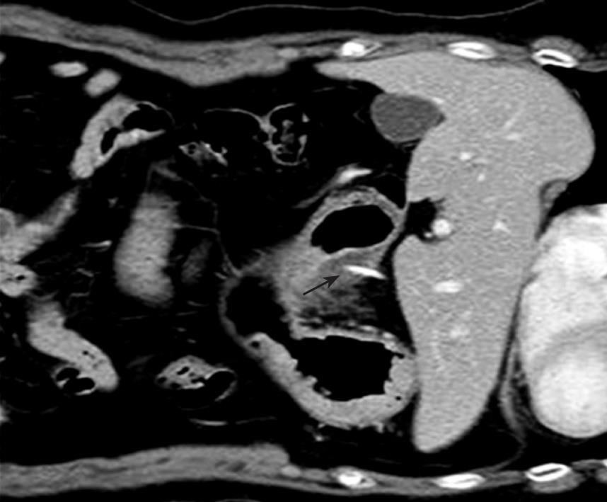
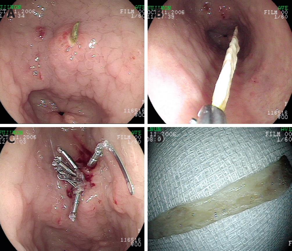
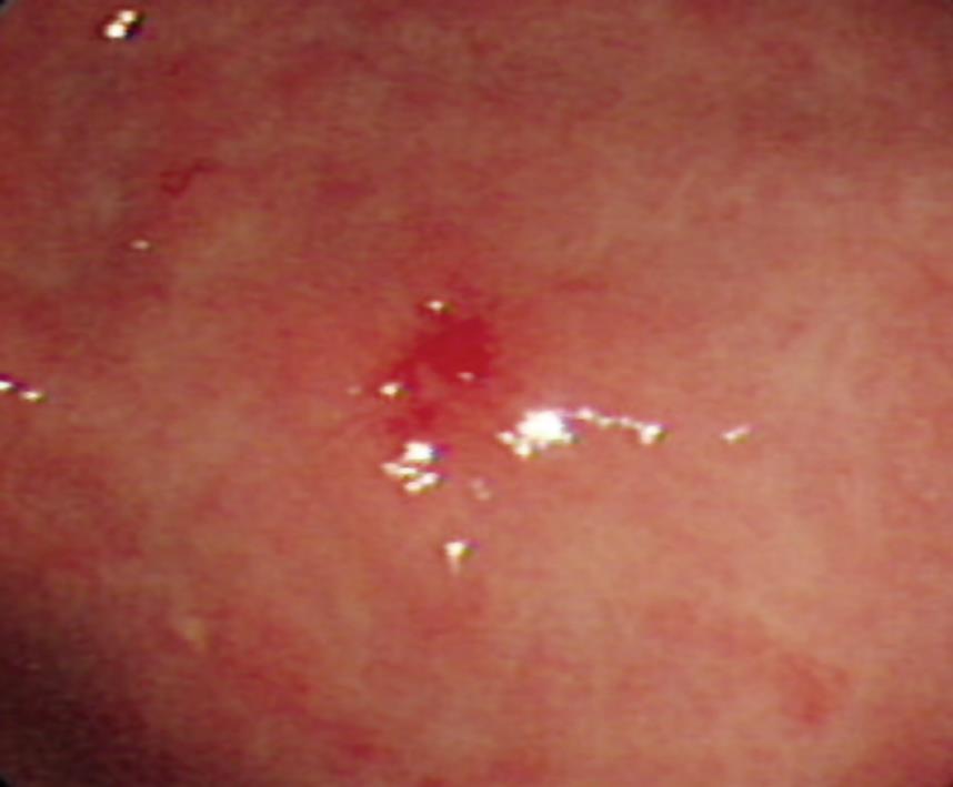
A 70-year old woman visited the emergency room complaining of epigastric pain for three days. She accidentally swallowed several lumps of poorly chewed chicken. She had a partially wearing denture because of teeth extraction one month ago. Physical examination disclosed mild tenderness in the periumbilical area without surgical signs including rebound tenderness. Her blood pressure was 100/60 mmHg, heart rate was 80/min, and body temperature was 36.6°C. The patient had an acute ill appearance but no history of nonsteroidal anti-inflammatory drug use, peptic ulcer, or liver disease. Laboratory studies revealed hemoglobin concentration of 132 g/L, hematocrit 38.6%, white blood cell count of 11.7 × 10⁹/L, and platelet count of 222 × 109/L. Her serum urea nitrogen concentration was 3.2 mmol/L, creatinine 50.4 &mgr;mol/L, AST 283 nkat/L, ALT 233 nkat/L, sodium 138 mmol/L, potassium 3.8 mmol/L, and chloride 104 mmol/L. Chest and abdomen X-ray images were negative for any abnormalities, but abdominal CT showed a suspicious penetration or perforation of the stomach wall measuring about 3 cm, by a linear radiopaque material at the lesser curvature of the antrum. The end of a chicken bone was very close to but did not penetrate the liver, and no major vessel injury was seen in the abdomen CT (Figure 1). Endoscopic examination revealed a chicken bone that penetrated into the prepyloric antrum (Figure 2A). The penetrating chicken bone was removed gently with grasping forceps (Figure 2B). No bleeding or other complications occurred after removal of the penetrating chicken bone. Five endoscopic clips were applied immediately at the removal site (Figure 2C) and the periumbilical pain resolved promptly. The removed bone fragment measured 3.0 cm in length (Figure 2D). Chest and flat abdomen X-ray imaging was performed serially after removal of the bone and revealed no abnormalities, such as free air. After removal of the bonegragment, the patient was treated with conservative care (nothing taken by mouth, intravenous hyper-alimentation, intravenous omeprazole, and antibiotics) for 3 d, after which she was completely asymptomatic and discharged without complication. Follow-up endoscopy was performed 7 d later and showed near closure of the ulcer at the penetration site (Figure 3).
Figure 1 2.
8 cm linear density (arrow) penetrating into the thickened gastric wall (CT).
Figure 2 Endoscopic management.
A: A chicken bone penetrating into the prepyloric antrum; B: The whole chicken bone was removed using a grasping forcep; C: Clip placement was performed at the removal site; D: The length of removed chicken bone was about 3.0 cm.
Figure 3 Follow- up endoscopy revealing near closure of the ulcer at the penetration site.
DISCUSSION
Foreign body ingestion and food bolus impaction occur commonly. Although most foreign bodies will pass out spontaneously, 10%-20% require nonoperative intervention, and 1% or fewer require surgical procedure[12]. The incidence of accidental chicken bone ingestion is 6%-6.4% in Asian countries[1213]. There are several reports on colon wall perforation by a chicken bone treated endoscopically. Tarnasky et al[14] have reported colonoscopic removal of a chicken bone impacted in the sigmoid colon without complication. Rex et al[5] have reported that two patients had their chicken bones impacted in the sigmoid removed successfully by colonoscopy. However, to our knowledge, no reports are available on patients with chicken bone penetration of the gastric wall treated endoscopically with clipping and conservative care.
In adults, foreign body ingestion occurs commonly among those with prison inmates, psychiatric patients, alcoholics, children, selected professions (carpenters and dressmakers), and people wearing dentures[15]. In our patient, the foreign body ingestion seemed to have resulted from poor dentition and an artificial denture.
Foreign bodies induce various clinical manifestations, such as perforation, bleeding, bowel obstruction, and even ureteral colic[1617]. A foreign body that perforates the bowel wall may take several possible courses, including lying in the bowel lumen at the site of perforation, like this patient, or passing through the gastrointestinal wall to migrate to a distal organ[18].
In the diagnosis of nonmetallic foreign bodies (especially fish or chicken bones), CT scanning is superior to plain X-ray radiography. Plain radiography is unreliable, even with bony radiopacity, because of the masking effect of the soft tissue mass, fluid collection around the penetrated bone, and the absence of free gas in the abdomen[619]. Also In this patient, CT scanning was a more reliable diagnostic method than X-ray. CT scanning showing the calcified foreign body with a thickened intestinal segment, localized pneumoperitoneum, regional fatty infiltration, and associated intestinal obstruction is definite for diagnosis of nonmetallic foreign body perforation[19]. However, the accuracy of CT is limited by the lack of observer awareness, and a high index of suspicion must be maintained for the correct diagnosis[20]. Immediate surgical intervention is a traditional treatment of choice for frank gastrointestinal perforation. However, it was reported in recent years, that endoscopic clip placement, as used in a hemostatic procedure, can be used in treatment of anastomotic leaks after esophagogastric surgery and gastrointestinal perforation[7–9]. Clipping has been used to treat foreign body removal (e.g., gastric toothpick penetration)[11]. We decided to perform endoscopic removal of the chicken bone penetrating into the gastric wall and to use the clip to close the penetration site because CT scanning showed no peritoneal irritation and no complication associated with the bone penetration. The pain and symptoms relating to the foreign body disappeared immediately after removal of the penetrating bone fragment. However, serious complications could have occurred if the penetrating bony fragment stuck to a vessel, or if a peritoneal abscess pocket resulted from the bone penetration. Thus, it is necessary to closely observe the patient’s status before and after such a procedure.
In conclusion, endoclipping can be a useful method to treat gastric penetration by foreign bodies, such as a chicken bone, in patients with no signs or symptoms of peritoneal irritation.


