INTRODUCTION
It is recognized that misidentification of normal anatomy, as well as the presence of anatomical variations, contributes to the occurrence of major postoperative complications, especially biliary injuries[1]. Such injuries can in turn cause significant morbidity and occasionally even mortality. Sound knowledge of the normal anatomy of the extrahepatic biliary tract, as well as the surgical implications[2], is thus essential to prevent these complications. Magnetic resonance cholangiopancreatography (MRCP) is a recently developed technique that allows non-invasive assessment of the biliary tree[3]. Anatomical variants of the cystic duct and cysticohepatic junction that may increase the risk of bile duct injury in biliary surgery are frequently identified with endoscopic retrograde cholangiopancreatography (ERCP), MRCP and percutaneous transhepatic cholangiography (PTC). We highlight two cases of anatomical variations of the cystic duct, in which the abnormality was found during surgery and subsequently confirmed by postoperative cholangiography and ERCP.
CASE REPORT
Case 1
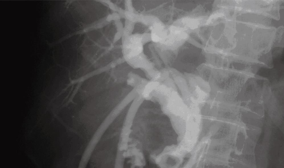
A 64-year-old man was admitted for surgery, with features of acute cholecystitis: right upper quadrant pain, vomiting and fever. Examination revealed a temperature of 38°C with tenderness in the right upper quadrant. He had previously been investigated for abdominal pain and had abdominal CT 4 years previously, which showed minimal intrahepatic ductal dilatation. Laboratory values showed a normal white cell count, total and direct biluribin was 24 &mgr;mol/L and 6 &mgr;mol/L respectively, alanine aminotransferase was 80 U/L, aspartate aminotransferase was 38 U/L, alkaline phosphatase was 301 U/L, and amylase was normal. Ultrasonography showed distension of the gallbladder with calculi and a thick wall associated with pericholecystic fluid. Cholelithiasis with chronic cholecystitis was diagnosed, and an open laparotomic cholecystectomy was planned. Upon laparatomy, the liver was normal and the gallbladder was thick-walled with minimal adhesion in Calot’s triangle and surrounding tissues. On separation, the entering of cystic was misidentified as being short and entering the right hepatic duct. The cystic duct was clipped and divided. The cystic artery was then identified, clipped and divided. Further exploration showed multiple calculi in the right and common hepatic ducts. Cholecystectomy was completed, followed by T-tube drainage of the common and right hepatic ducts (for postoperative confirmation and documentation). The surgical procedure was followed by abdominal pain, and T-tube cholangiography was requested on postoperative d 14. Cholangiography through a T-tube showed that the cystic and right hepatic ducts were comparatively long. Long cystic duct with low medial insertion into common bile duct was made (Figure 1). The entire bile duct was identified and found to be intact. Six weeks later, the retained stones was extracted by cholangioscopy through the sinus tract of the T-tube. The patient was successfully cured.
Figure 1 The cystic and left-right hepatic ducts were comparatively long, and the cystic duct opening was located in the distal choledochus with retained stones.
Two T tubes were respectively located in the cystic duct and in the common hepatic duct.
Case 2
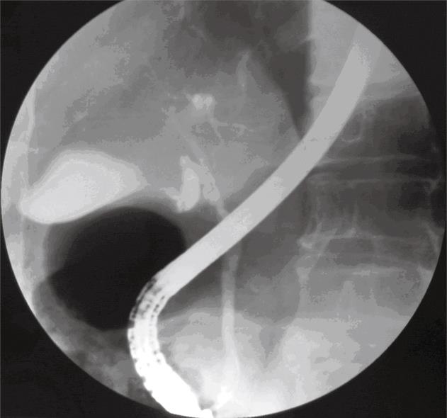
A 41-year-old woman patient presented with 3-year discontinuous right upper quadrant pain that radiated into her back in keeping with biliary colic, sometimes accompanied with fever and nausea. The pain was aggravated with greasy food. In the course of the present illness, she had received medication for acid peptic disease and parasitic infestations, with no improvement. She had a similar history in the past. Physical examination revealed a soft, non-distended abdomen and no tenderness in the right upper quadrant, without peritoneal signs. Murphy’s sign was negative. Her temperature was 37.5°C, and the rest of her vital signs were normal. Laboratory analysis of liver function was normal. US revealed no abnormalities. The patient was observed at rest with continuous gastrointestinal decompression and fluid infusion. Her symptoms improved significantly within 4 d of treatment. However, a few days later, she experienced right upper quadrant pain. ERCP was requested and revealed a long cystic duct with a narrow and in-curved lumen, which was well separated from the gallbladder. The rest of the entire biliary tract was normal without calculi (Figure 2). A narrow-winding cystic duct was created, followed by cholecystectomy. The surgical procedure was followed by an uneventful convalescence until discharge.
Figure 2 ERCP showing a narrow-winding cystic duct.
DISCUSSION
Anatomical variations of the cystic duct are usually of no clinical significance, occurring in 18%-23% of cases[4]. However, unrecognized variant anatomy can be a source of confusion on imaging studies. In addition, the cystic duct may be involved in a wide variety of both primary and secondary disease processes. The rate of injury varies in the medical literature from 0% to 1%[5]. The following are some of the cystic duct variations found: (1) the cystic and common hepatic duct are in parallel; (2) low confluence of the cystic duct[2]; (3) insertion of the cystic duct in the left and right hepatic ducts, and bifurcation of the left and right hepatic ducts[2]; (4) anterior, posterior spiral types of insertion of the cystic duct on the left side of the common hepatic duct; (5) parahepatic duct insertion into the cystic duct; (6) absent or short cystic duct (length < 5 mm); (7) cystic duct hypertrophy, with a diameter > 5 mm; (8) double cystic duct[67]; (9) right hepatic duct emptying into the cystic duct[8]; and (10) hepaticocystic duct[9], a very rare congenital abnormality in which the common hepatic duct enters the gallbladder. The left, right, and common hepatic ducts are all defective, with the cystic duct draining the entire biliary system into the duodenum.
Multiple modalities permit depiction of the normal anatomy, as well as disease processes of the cystic duct, including CT, PTC, ERCP, intraoperative cholangiography and MRCP. Although visualization of the dilated cystic duct is possible with US and CT, the normal-caliber cystic duct may be difficult to detect with these techniques[10]. In our first case, CT demonstrated minimal intrahepatic ductal dilatation, but failed to show low insertion of the cystic duct, as was revealed by surgery. In this case, the low insertion of the cystic duct was misdiagnosed as gallbladder and bile duct calculi. However, in the second case, ERCP showed a long cystic duct with a narrow and in-curved lumen. An anomalous cystic duct was diagnosed before surgery. Anatomical variation is readily identified at ERCP. In clinical practice, if the patient presents with intermittent non-colic right upper abdominal pain, and ultrasound, CT and endoscopy eliminate choledocholithiasis, tumor and peptic ulcer, then a narrow-winding cystic duct should be considered. ERCP is extremely helpful in diagnosis. Recent studies have demonstrated that MRCP may provide a non-invasive alternative to ERCP and PTC in diagnosis of anomalous cystic ducts[11]. Taourel and colleagues[12] evaluated the accuracy of MRCP in the diagnosis of anatomic variants of the biliary tree in 171 patients. MRCP demonstrated a cystic duct in 126 patients (74%), including low cystic duct insertion in 11 (9%) and a parallel course of the cystic and hepatic ducts in 31 patients (25%). These findings suggest that accurate preoperative assessment is very useful in providing a surgical treatment plan in addition to confirming diagnosis. During cholecystectomy, to avoid biliary tree injury, it is important to identify the common hepatic-cystic duct junction. Misidentification of the cystic duct can lead to postoperative complications. In particular, attention should pay to low medial insertion of the cystic duct because this anatomical variant may lead to misdiagnosis on imaging, and thus affect therapeutic intervention, as was seen in our first case.
A limited literature review of bile duct variation has shown that the aim of most surgeons is to identify whether there are bile duct stones. With respect to the accidental discovery of bile duct variation, it is not the nature of the variation itself but rather the existence of the bile duct variation that is the most important factor in the prevention of bile duct injury. Most injuries to the cystic duct usually occur when it runs parallel to the common bile duct and is encased in a common sheath, so that separation between the ducts is not readily apparent at surgery. T-tube placement in the cystic duct remnant is usually of no consequence; however, there may be a difficulty if retained common duct stones are present, and stone removal via the T-tube is attempted. In such cases, access to the bile duct is via a tract that enters the cystic duct, and manipulation and extraction must occur via the cystic duct across the valves of Heister. Stone extraction is more difficult or may be impossible via this route[13]. Suspicion should be raised if the cystic duct is of an unusually large calibre. Intraoperative cholangiography should be used in case of doubt and, in unusual circumstances, cholangiography can be performed via the gallbladder to aid in the identification of the cystic duct as well as the common bile duct.
In conclusion, the cystic duct may be involved in a variety of anatomical variations. Diagnostic accuracy relies on a clear understanding of the normal anatomy and anatomical variants of the cystic duct, and imaging features of calculous disease.


