Published online Nov 7, 2007. doi: 10.3748/wjg.v13.i41.5525
Revised: July 7, 2007
Accepted: August 25, 2007
Published online: November 7, 2007
There are occasional incidences of gallstone spillage during laparoscopic cholecystectomy, and there have been frequent reports on such a topic in the literature. To the best of our knowledge, however, there have been no reports about spilled stones caused by spontaneously perforated hemorrhagic cholecystitis. Here, we report the radiologic findings of spilled stones caused by spontaneously perforated hemorrhagic cholecystitis in a 55-year-old man.
- Citation: Kim YC, Park MS, Chung YE, Lim JS, Kim MJ, Kim KW. Gallstone spillage caused by spontaneously perforated hemorrhagic cholecystitis. World J Gastroenterol 2007; 13(41): 5525-5526
- URL: https://www.wjgnet.com/1007-9327/full/v13/i41/5525.htm
- DOI: https://dx.doi.org/10.3748/wjg.v13.i41.5525
With the increased use of laparoscopic surgery, the spillage of gallstones during laparoscopic cholecystectomy has been reported in 6%-40% of cases[1,2]. To the best of our knowledge, however, there have been no reports about spilled stones caused by spontaneously perforated hemorrhagic cholecystitis. Here, we present ultrasonography (US) and computed tomography (CT) images of this rare condition.
A 55-year-old man complained of abrupt upper abdominal pain during hospitalization for a brain abscess. A complete blood count taken 12 h after the attack showed that the level of hemoglobin dropped to 8.8 g/L (from 13.3 g/L, 36 h before the attack). Other blood analysis revealed mild thrombocytopenia (platelet count 106 × 103/μL), and mild hyperbilirubinemia (total bilirubin concentration 1.7 mg/dL); however, the white blood cell count was normal (9.27 × 10⁶/μL).
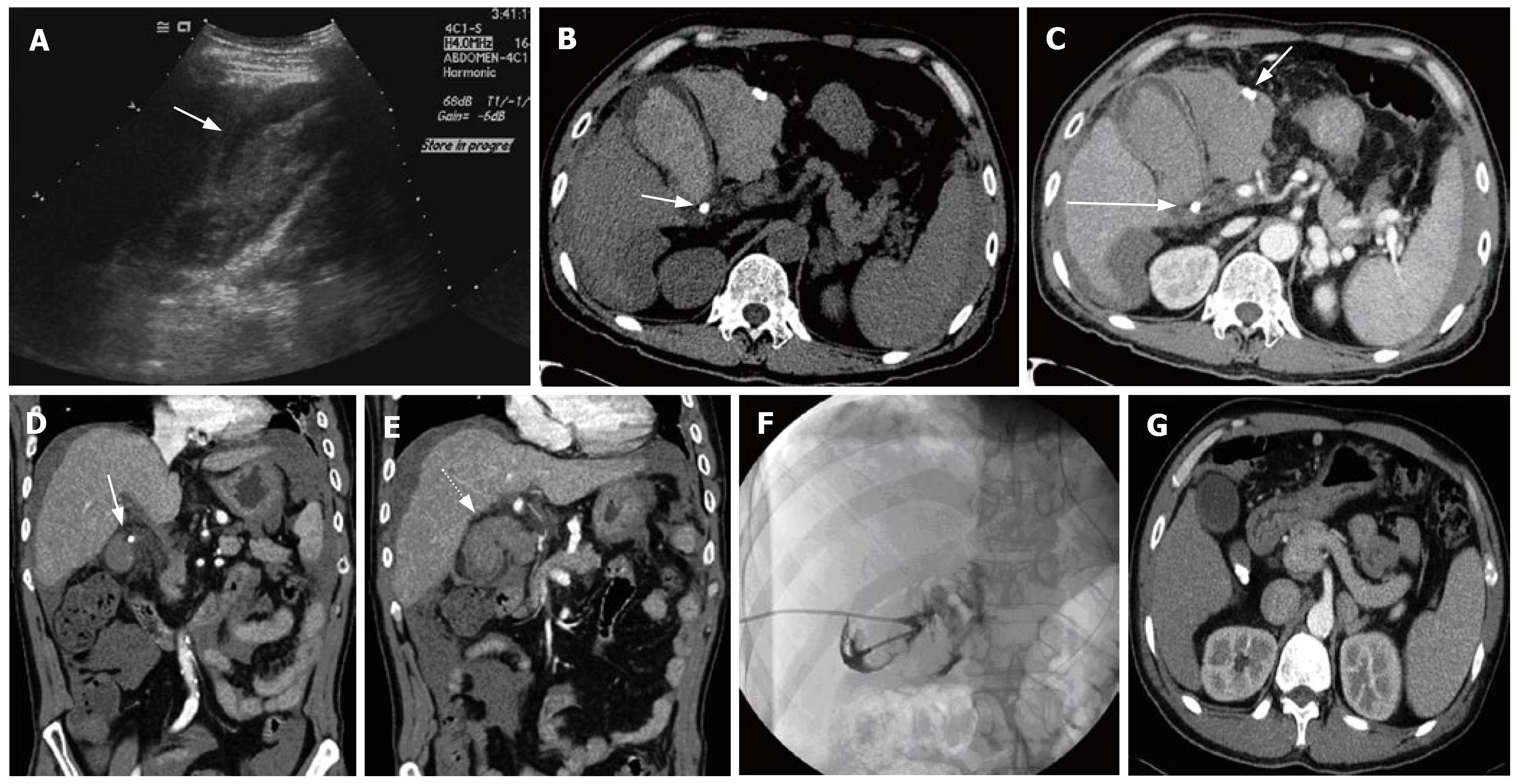
Immediately after the attack, the patient underwent US, which demonstrated echogenic material in the gallbladder lumen (Figure 1A), with a positive sonographic Murphy's sign. US was discontinued because the patient complained of severe abdominal pain. Contrast-enhanced CT was performed and its images revealed high-density fluid, both inside and outside the gallbladder. One impacted cystic duct stone was seen, as were several calcified objects (which looked like stones), within the high-density (46-61 HU) fluid surrounding the gallbladder (Figure 1B and C). A defect in the wall or mucosal disruption of the gallbladder was also noted (Figure 1D and E). In addition, underlying liver cirrhosis with splenomegaly was observed. Percutaneous transhepatic gallbladder drainage (PTGBD) and cholangiography were performed. Cholecystography demonstrated contrast leakage from the gallbladder (Figure 1F).
He had a medical history of several years of alcoholic liver cirrhosis with mild esophageal varices and multiple gallbladder stones. Two months before the current attack, he underwent an abdominal CT scan to evaluate liver cirrhosis. At that time, CT revealed multiple stones in the gallbladder, without complications (Figure 1G).
Laparoscopic cholecystectomy has become a popular alternative to open surgery for the treatment of gallstones. With the increase in laparoscopic cholecystectomy, the incidence of gallstone spillage has increased, with an incidence ranging from 6% to 40%[1,2]. The complications of peritoneal spilled gallstones are abscess, fistula formation within various intraperitoneal organs, or sinus tract formation[3-7]. To the best of our knowledge, however, gallstone spillage caused by spontaneously perforated hemorrhagic cholecystitis in a patient who did not undergo cholecystectomy has not been reported.
The proposed mechanism of gallbladder perforation is stone impaction in the cystic duct, which leads to retention of secretion from mucus glands and distention, with progressive distention leading to vascular compromise, followed by necrosis and perforation[8]. During this process, bleeding can occur, which results in hemorrhagic cholecystitis with hemoperitoneum. In our case, the patient had liver cirrhosis and therefore the risk of bleeding could have been increased.
In our study, US showed heterogeneous, highly echogenic material, both within and outside the gallbladder lumen, which may have been suggestive of gallbladder perforation with hemorrhage. However, we could not detect the exact perforation site nor spilled gallstones on US, and so we were not able to diagnose gallbladder perforation at that time. CT clearly demonstrated the perforation site at the gallbladder wall and spilled radiopaque stones that were missed on US. It has been reported that distended gallbladder, thickened and bulging gallbladder wall, pericholecystic fluid, cholelethiasis, and gallbladder wall defects are the US and CT findings of gallbladder perforation. The most specific of these findings is gallbladder wall defects, with a detection rate of 38.4% on US and 69.2% on CT[9]. The most common site of perforation is reported to be the fundus (70% of cases), due to poor vascular supply[10].
In conclusion, gallstone spillage due to spontaneous perforation is a very rare condition. However, US or CT visualization of mucosal disruption of the gallbladder wall, with gallstones within hemoperitoneum is suggestive of the condition.
S- Editor Liu Y L- Editor Kerr C E-Editor Li JL
| 1. | Fletcher DR, Hobbs MS, Tan P, Valinsky LJ, Hockey RL, Pikora TJ, Knuiman MW, Sheiner HJ, Edis A. Complications of cholecystectomy: risks of the laparoscopic approach and protective effects of operative cholangiography: a population-based study. Ann Surg. 1999;229:449-457. [RCA] [PubMed] [DOI] [Full Text] [Cited by in Crossref: 355] [Cited by in RCA: 327] [Article Influence: 12.6] [Reference Citation Analysis (0)] |
| 2. | Ahmad SA, Schuricht AL, Azurin DJ, Arroyo LR, Paskin DL, Bar AH, Kirkland ML. Complications of laparoscopic cholecystectomy: the experience of a university-affiliated teaching hospital. J Laparoendosc Adv Surg Tech A. 1997;7:29-35. [RCA] [PubMed] [DOI] [Full Text] [Cited by in Crossref: 33] [Cited by in RCA: 33] [Article Influence: 1.2] [Reference Citation Analysis (0)] |
| 3. | Woodfield JC, Rodgers M, Windsor JA. Peritoneal gallstones following laparoscopic cholecystectomy: incidence, complications, and management. Surg Endosc. 2004;18:1200-1207. [RCA] [PubMed] [DOI] [Full Text] [Cited by in Crossref: 98] [Cited by in RCA: 114] [Article Influence: 5.4] [Reference Citation Analysis (0)] |
| 4. | Diez J, Arozamena C, Gutierrez L, Bracco J, Mon A, Sanchez Almeyra R, Secchi M. Lost stones during laparoscopic cholecystectomy. HPB Surg. 1998;11:105-108; discuss 108-109. [PubMed] |
| 5. | Hui TT, Giurgiu DI, Margulies DR, Takagi S, Iida A, Phillips EH. Iatrogenic gallbladder perforation during laparoscopic cholecystectomy: etiology and sequelae. Am Surg. 1999;65:944-948. [PubMed] |
| 6. | Rice DC, Memon MA, Jamison RL, Agnessi T, Ilstrup D, Bannon MB, Farnell MB, Grant CS, Sarr MG, Thompson GB. Long-term consequences of intraoperative spillage of bile and gallstones during laparoscopic cholecystectomy. J Gastrointest Surg. 1997;1:85-90; discussion 90-1. [RCA] [PubMed] [DOI] [Full Text] [Cited by in Crossref: 76] [Cited by in RCA: 77] [Article Influence: 2.8] [Reference Citation Analysis (0)] |
| 7. | Schäfer M, Suter C, Klaiber C, Wehrli H, Frei E, Krähenbühl L. Spilled gallstones after laparoscopic cholecystectomy. A relevant problem? A retrospective analysis of 10,174 laparoscopic cholecystectomies. Surg Endosc. 1998;12:305-309. [RCA] [PubMed] [DOI] [Full Text] [Cited by in Crossref: 88] [Cited by in RCA: 87] [Article Influence: 3.2] [Reference Citation Analysis (0)] |
| 8. | Madrazo BL, Francis I, Hricak H, Sandler MA, Hudak S, Gitschlag K. Sonographic findings in perforation of the gallbladder. AJR Am J Roentgenol. 1982;139:491-496. [RCA] [PubMed] [DOI] [Full Text] [Cited by in Crossref: 57] [Cited by in RCA: 48] [Article Influence: 1.1] [Reference Citation Analysis (0)] |
| 9. | Kim PN, Lee KS, Kim IY, Bae WK, Lee BH. Gallbladder perforation: comparison of US findings with CT. Abdom Imaging. 1994;19:239-242. [RCA] [PubMed] [DOI] [Full Text] [Cited by in Crossref: 82] [Cited by in RCA: 71] [Article Influence: 2.3] [Reference Citation Analysis (0)] |
| 10. | Massie JR, Coxe JW, Parker C, Dietrick R. Gall bladder perforations in acute cholecystitis. Ann Surg. 1957;145:825-831. [RCA] [PubMed] [DOI] [Full Text] [Cited by in Crossref: 10] [Cited by in RCA: 11] [Article Influence: 0.2] [Reference Citation Analysis (0)] |


