Published online Jul 14, 2007. doi: 10.3748/wjg.v13.i26.3631
Revised: March 4, 2007
Accepted: April 18, 2007
Published online: July 14, 2007
We report a case of right lower abdominal wall and groin abscess resulting from acute appendicitis. The patient was an 27-year-old man who had no apparent abdominal signs and was brought to the hospital due to progressive painful swelling of right lower abdomen and the groin for 10 d. Significant inflammatory changes of soft tissue involving the right lower trunk were noted without any apparent signs of peritonitis. Laboratory results revealed leukocytosis. Abdominal ultrasonography described the presence of abscess at right inguinal site also communicating with the intraabdominal region. Right inguinal exploration and laparotomy were performed and about 250 mL of pus was drained from the subcutaneous tissue and preperitoneal space. No collection of pus was found intraabdominally and subserous acute appendicitis was the cause of the abscess. The patient fully recovered at the end of the second post-operation week. This case reminds us that acute appendicitis may have an atypical clinical presentation and should be treated carefully on an emergency basis to avoid serious complications.
- Citation: Yildiz M, Karakayali AS, Ozer S, Ozer H, Demir A, Kaptanoglu B. Acute appendicitis presenting with abdominal wall and right groin abscess: A case report. World J Gastroenterol 2007; 13(26): 3631-3633
- URL: https://www.wjgnet.com/1007-9327/full/v13/i26/3631.htm
- DOI: https://dx.doi.org/10.3748/wjg.v13.i26.3631
Acute appendicitis is considered one of the elementals of general surgical disease processes, yet its presentation often confounds its diagnosis by most surgeons. Its presentation as abscess in the abdominal wall and groin is a rare clinical entity. Because of insidious onset and subtle clinical signs of resulting abscess, the diagnosis of such cases is often delayed[1,2]. Acute appendicitis such as those forming extensive abscesses may sometimes become complicated and require a prolonged treatment period[3,4]. These complications should be kept in mind in order to avoid further sequelae. We hereby present a rare case of unruptured acute appendicitis with extensive formation of right lower abdominal wall and groin abscess. We aim to emphasize the importance of suspecting certain intra-abdominal inflammatory conditions in the differential diagnosis, early diagnosis and prompt management for this disease.
A 27-year-old man presented with pain for 10 d in the lower abdomen and groin. He also experienced a swollen and red groin and painful disability of the right thigh.His appetite was normal initially, but lessened for the last a few days. The patient denied any other symptoms of gastrointestinal dysfunction. On physical examination, the patient appeared ill with a heart rate of 96/min, respiratory rate of 28/min, blood pressure of 110/80 mmHg and body temperature of 38.6°C.
There were significant inflammatory signs such as local heat, swelling,edema and tenderness involving the entire right abdominal wall, especially disclosing the right groin area. No subcutaneous emphysema or crepitation was noted. The abdominal examination revealed unremarkable signs during palpation and the peritoneal reaction was absent.
Laboratory data showed leukocytosis with a WBC count of 25.700/mm3, of which 80% were neutrophils. All the other blood chemical tests were within the normal range. Chest and abdominal X-ray revealed no abnormality. An ultrasound scan suggested a fluid collection in the right inguinal region communicating with the abdomen, which was evaluated to be an abscess.
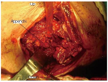
The clinical, radiological and laboratory findings showed the presence of inguinal abscess as the source of sepsis. The abscess in the groin was opened through an inguinal incision. The subcutaneous and preperitoneal tissues were affected by purulent material. The abscess was found between the muscle groups and dissected downward to the right thigh. As the destroyed muscle layers entered towards the peritoneum, the tip of the appendix was protruding through the incision (Figure 1). The abdomen was opened immediately and the exploration revealed an subserous acutely inflamed appendix. However, the intra-abdominal cavity was clear without any contamination. Appendectomy was done and the debridement of the non-viable abdominal wall muscle groups and the drainage of the abscess cavity were performed. The bacterial culture revealed Escherichia coli and Klebsiella pneumonia infection. The patient began to take oral fluids in 2 d and recovered smoothly after surgery. He was discharged uneventfully 2 wk after the surgery.
A review of the literature suggests certain intra-abdominal inflammatory pathologies in the etiology of painful, swollen groin and thigh, such as diverticulitis, acute appendicitis, Crohn's disease, colorectal carcinoma, rectal trauma and primary staphylococcal abscess[1,2,4]. But surprisingly, acute appendicitis, although among the most common surgical emergencies, has rarely been discussed with such unusual presentation. The etiopathogenesis of this presentation in this case can be explained by the direct contamination of the right anterior abdominal wall and groin by an inflamed phlegmonous appendix.The spread of resultant sepsis along the abdominal wall muscles, preperitoneal space and downward behind the inguinal ligament into the thigh thus presented clinically as an abscess[1,2,5].
In many cases, the previous history and presenting symptoms did not clearly indicate an intra-abdominal origin, and this has often created serious diagnostic problems. Admitting diagnosis of inguinal abscess could not be established until surgery provided a definitive diagnosis. Negative appendectomy rates have been relatively stable over the years and all patients are affected to one extent or another[4,6,7]. Currently, there is no test or objective physical finding which can rule out the presence of appendicitis with acceptable accuracy.
The insidious onset of abscess formation may not be responsive to medical treatment. Only the results of bacterial culture if could be obtained before surgery, could indicate the nature of the abscess. The most common causative pathogen of primary abscess is staphylococcus aureus, while that of secondary abscess is usually mixed intestinal floras[8-10]. In this case, bacterial examination revealed the organisms Escherichia coli and Klebsiella pneumonia which suggested the intestinal relation. In a study by Hale et al, intraoperative cultures of peritoneal fluid were obtained in 2522 cases. The most commonly isolated organism was Escherichia coli followed by Enterococcus and other Streptoccus species. Less commonly encountered organisms included Pseudomonas, Klebsiella and Bacteriodes species[4].
In most of the reported cases, a computed tomographic scan (CT) is used as a modality for definite diagnosis. Abdominal CT scan helps not only establish the diagnosis but also evaluates the extension of involvement[4,11-13]. Abdominal ultrasonography is also a modality widely used for diagnosis of abdominal pathologies. It is also helpful in the evaluation of its treatment. In the present case, ultrasonography suggested the relationship with the abdomen and guided us to the intra-abdominal origin of the abscess. A CT imaging could not be performed in this case because of technical problems.
The drainage of abscess can be achieved by percu-taneous approach or by laparotomy based on US and CT findings. There are several reports demonstrating substantial results by percutaneous drainage of the abscess and then by surgery only if percutaneous drainage fails[8,14,15]. Percutaneous approach would probably be not adequate for this case, because our patient was in a critical condition requiring prompt drainage which can not be achieved by percutaneous method and the infection could not be controlled. Surgery seems to be advantageous over percutaneous drainage in patients under critical condition such as appendicitis, perforated appendicitis, diverticulitis or malignancy[8].
A search for the presence of intra-abdominal pathology by a thorough clinical and radiological evaluation should be made in all patients with unexplained groin and thigh symptoms with fever and leukocytosis. Clinicians should be aware that an abscess may be the manifestation of an intestinal disorder despite minimal abdominal signs and to improve the survival rate, the clinical diagnosis must be pursued aggressively.
S- Editor Liu Y L- Editor Ma JY E- Editor Lu W
| 1. | Sharma SB, Gupta V, Sharma SC. Acute appendicitis presenting as thigh abscess in a child: a case report. Pediatr Surg Int. 2005;21:298-300. [RCA] [PubMed] [DOI] [Full Text] [Cited by in Crossref: 16] [Cited by in RCA: 18] [Article Influence: 0.9] [Reference Citation Analysis (0)] |
| 2. | El-Masry NS, Theodorou NA. Retroperitoneal perforation of the appendix presenting as right thigh abscess. Int Surg. 2002;87:61-64. [PubMed] |
| 3. | Lally KP, Cox CS, Androssy RJ. Appendix. Sabiston textbook of surgery. 17th ed. philadephia: Elsevier Saunders 2004; 1381-1399. |
| 4. | Hale DA, Molloy M, Pearl RH, Schutt DC, Jaques DP. Appendectomy: a contemporary appraisal. Ann Surg. 1997;225:252-261. [RCA] [PubMed] [DOI] [Full Text] [Cited by in Crossref: 218] [Cited by in RCA: 187] [Article Influence: 6.7] [Reference Citation Analysis (0)] |
| 5. | Jager GJ, Rijssen HV, Lamers JJ. Subcutaneous emphysema of the lower extremity of abdominal origin. Gastrointest Radiol. 1990;15:253-258. [RCA] [PubMed] [DOI] [Full Text] [Cited by in Crossref: 21] [Cited by in RCA: 23] [Article Influence: 0.7] [Reference Citation Analysis (0)] |
| 6. | Edwards JD, Eckhauser FE. Retroperitoneal perforation of the appendix presenting as subcutaneous emphysema of the thigh. Dis Colon Rectum. 1986;29:456-458. [RCA] [PubMed] [DOI] [Full Text] [Cited by in Crossref: 23] [Cited by in RCA: 25] [Article Influence: 0.6] [Reference Citation Analysis (0)] |
| 7. | Jones PF. The influence of age and gender on normal appendicectomy rates. Aust N Z J Surg. 1988;58:919-920. [RCA] [PubMed] [DOI] [Full Text] [Cited by in Crossref: 2] [Cited by in RCA: 3] [Article Influence: 0.1] [Reference Citation Analysis (0)] |
| 8. | Hsieh CH, Wang YC, Yang HR, Chung PK, Jeng LB, Chen RJ. Extensive retroperitoneal and right thigh abscess in a patient with ruptured retrocecal appendicitis: an extremely fulminant form of a common disease. World J Gastroenterol. 2006;12:496-499. [PubMed] |
| 9. | Rotstein OD, Pruett TL, Simmons RL. Thigh abscess. An uncommon presentation of intraabdominal sepsis. Am J Surg. 1986;151:414-418. [RCA] [PubMed] [DOI] [Full Text] [Cited by in Crossref: 34] [Cited by in RCA: 34] [Article Influence: 0.9] [Reference Citation Analysis (0)] |
| 10. | Ricci MA, Rose FB, Meyer KK. Pyogenic psoas abscess: worldwide variations in etiology. World J Surg. 1986;10:834-843. [RCA] [PubMed] [DOI] [Full Text] [Cited by in Crossref: 199] [Cited by in RCA: 211] [Article Influence: 5.4] [Reference Citation Analysis (0)] |
| 11. | Haaga JR. Imaging intraabdominal abscesses and nonoperative drainage procedures. World J Surg. 1990;14:204-209. [RCA] [PubMed] [DOI] [Full Text] [Cited by in Crossref: 26] [Cited by in RCA: 29] [Article Influence: 0.8] [Reference Citation Analysis (0)] |
| 12. | Ishigami K, Khanna G, Samuel I, Dahmoush L, Sato Y. Gas-forming abdominal wall abscess: unusual manifestation of perforated retroperitoneal appendicitis extending through the superior lumbar triangle. Emerg Radiol. 2004;10:207-209. [RCA] [PubMed] [DOI] [Full Text] [Cited by in Crossref: 16] [Cited by in RCA: 15] [Article Influence: 0.7] [Reference Citation Analysis (0)] |
| 13. | Kim S, Lim HK, Lee JY, Lee J, Kim MJ, Lee AS. Ascending retrocecal appendicitis: clinical and computed tomographic findings. J Comput Assist Tomogr. 2002;30:772-776. [RCA] [PubMed] [DOI] [Full Text] [Cited by in Crossref: 25] [Cited by in RCA: 29] [Article Influence: 1.5] [Reference Citation Analysis (0)] |
| 14. | Benoist S, Panis Y, Pannegeon V, Soyer P, Watrin T, Boudiaf M, Valleur P. Can failure of percutaneous drainage of postoperative abdominal abscesses be predicted? Am J Surg. 2002;184:148-153. [RCA] [PubMed] [DOI] [Full Text] [Cited by in Crossref: 65] [Cited by in RCA: 69] [Article Influence: 3.0] [Reference Citation Analysis (0)] |
| 15. | Cantasdemir M, Kara B, Cebi D, Selcuk ND, Numan F. Computed tomography-guided percutaneous catheter drainage of primary and secondary iliopsoas abscesses. Clin Radiol. 2003;58:811-815. [RCA] [PubMed] [DOI] [Full Text] [Cited by in Crossref: 57] [Cited by in RCA: 61] [Article Influence: 2.8] [Reference Citation Analysis (0)] |


