Case Report Open Access
Copyright ©2007 Baishideng Publishing Group Co., Limited. All rights reserved.
World J Gastroenterol. Jun 21, 2007; 13(23): 3265-3267
Published online Jun 21, 2007. doi: 10.3748/wjg.v13.i23.3265
Gastrointestinal stromal tumor of appendix incidentally diagnosed by appendiceal hemorrhage
Kyu-Jong Kim, Won Moon, Moo In Park, Seun Ja Park, Department of Internal Medicine, Kosin University College of Medicine, Busan, South Korea
Seung Hyun Lee, Department of General Surgery, Kosin University College of Medicine, Busan, South Korea
Bong Kwon Chun, Department of Pathology, Kosin University College of Medicine, Busan, South Korea
Author contributions: All authors contributed equally to the work.
Correspondence to: Kyu-Jong Kim, MD, Division of Gastro-enterology, Kosin University College of Medicine, 34 Amnam-dong, Seo-gu, Busan 602-702, South Korea. drkkj@paran.com
Telephone: +82-51-9905206 Fax: +82-51-9905206
Received: March 21, 2007
Revised: April 6, 2007
Accepted: April 16, 2007
Published online: June 21, 2007

Abstract

Gastrointestinal stromal tumor is rare, which arises from the mesenchymal tissues in the gastrointestinal tract, and it is extremely rare in the appendix. Only a few cases have been found in this location to date. Although the annual incidence of lower gastrointestinal bleeding has been increasing, bleeding related to the appendix is quite rare. We herein present a very rare case of gastrointestinal stromal tumor incidentally found by appendiceal hemorrhage.

Key Words: Gastrointestinal stromal tumor; Gastrointestinal tract; Appendix; Lower gastrointestinal bleeding; Appendiceal hemorrhage


INTRODUCTION

The specific causes of lower gastrointestinal bleeding (LGIB) identified include colonic diverticula, angiodys-plasia, hemorrhoids, NSAID-induced injury, colonic ischemia, inflammatory bowel disease, and post-polypectomy complications[1]. Bleeding related to the appendix, although the annual incidence of LGIB has been increasing to date, is extremely rare, only a small number of reports have been documented.

Mesenchymal tumors of the appendix are scarce, and of them, few cases of gastrointestinal stromal tumor (GIST) have been found. Some of the tumors previously defined as appendiceal smooth muscle tumors might have been GIST because immunohistochemical studies in detail had not been evaluated in those cases; nevertheless, few cases have been reported so far[2,3].

We report a unique case of appendiceal GIST in-cidentally diagnosed based on the active LGIB occurring in the appendiceal mucosal defect of indefinite cause.

CASE REPORT

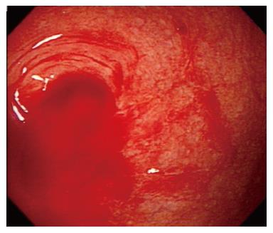
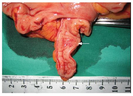
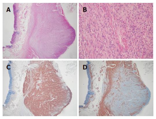
A 56-year-old man was admitted to our department on an emergency basis because of a sudden onset of hematochezia. He took a few tablets of NSAID to control the headache the night before. He denied abdominal pain and prior episodes of diarrhea and melena. He had no significant medical background and family history. The patient had a resting tachycardia. But, his blood pressure was normal. No abnormal findings were revealed throughout physical examination and laboratory tests except slightly decreased hemoglobin (11.6 g/dL). One unit of blood was immediately infused, and an urgent colonoscopy was performed without bowel preparation. A large amount of fresh blood was present along the entire colon, but not terminal ileum. In the cecum, an oozing of fresh blood from the appendiceal orifice was identified and it continued irrespective of repeated perendoscopic vigorous irrigation (Figure 1). Endoscopic hemostasis was not attempted due to no evidence of ulcerative lesions including any exposed vessel within or adjacent to the orifice. Multiple diverticulas of various sizes without evidence of active bleeding were also found in the proximal portion of the ascending colon and cecum (Figure 2). Surgical management for the arrest of continuous hemorrhage was performed. Right hemicolectomy with appendectomy was performed because the diverticulas around the appendiceal orifice as the focus of the bleeding could not be excluded absolutely. Macroscopically, the mucosa of the appendiceal specimen was normal at the time of surgery. But, a small nodule was found in the mid portion of the appendix (Figure 3). Microscopically, an ill-defined mass in the appendix which is composed of spindle cells of fascicular pattern, involving the proper muscle showed immunopositive staining for CD34, but not in smooth muscle actin, S-100, and c-KIT (Figure 4 A-D). Since the operation, the patient has experienced no further episodes of gastrointestinal bleeding for 3 mo.

Figure 1
Figure 1 Continuous oozing of fresh blood is seen from the appendiceal orifice.
Figure 2
Figure 2 Small diverticula without evidence of active bleeding are found in the cecum.
Figure 3
Figure 3 The inside of the appendiceal specimen appears normal, but a small nodule is found in the mid portion (white arrow).
Figure 4
Figure 4 A: Whole layers of appendix show an ill-defined mass involving the proper muscle layer (HE, × 20); B: The mass is composed of spindle cells usually in fascicular pattern. The neoplastic cells are characterized by spindle nuclei with blunt or tapered end and bland chromatin pattern and by various amounts of amphophilic cytoplasm and ill-defined cellular borders (HE, × 200); C: The neoplastic cells show immunopositive staining for CD34 (Immunohistochemical stain, × 20); D: The neoplastic cells show immunonegative staining for smooth muscle actin, but positive in the remaining proper muscle cells (Immunohistochemical stain, × 20).
DISCUSSION

This is an interesting case because the lower gastrointestinal bleeding (LGIB) was observed in appendiceal orifice, and GIST was found incidentally in surgery which was performed for the control of the bleeding.

LGIB, which is defined as bleeding beyond the ligament of Treitz, represents a diverse range of bleeding sources and severities. Its annual incidence of hospitalization was estimated to be 20-30 per 100 000 persons per year with increasing frequency, particularly in the elderly, it is approximately one-fifth as common as upper gastro-intestinal bleeding[4]. The sources of LGIB identified are diverticular bleeding, angiodysplasia, hemorrhoids, NSAID-induced, colonic ischemia, inflammatory bowel disease, post-polypectomy bleeding, infectious colitis, radiation colitis, neoplasms, stercoral ulcers, and others included bleeding from the appendiceal orifice by the less common causes[1].

This case is a patient with LGIB occurring in the appendix. Bleeding from the appendix with any cause, especially identified as the direct visualization at the time of colonoscopy is quite rare. To our knowledge with extensive reviews, a small number of reports have been available in the literature so far. The final diagnoses of these reports are listed as mucocele[5], diverticulum[6], aorto-appendiceal fistula[7-9], endometriosis[10], vascular malformation[11,12], transmural inflammation without evidence of well-formed granulomas[13], intussusception[14], isolated appendiceal Crohn’s disease[15-18], and aspirin-induced ulcer[19]. In this case, LGIB was related to NSAID-induced erosion with abundant RBC in the appendix near the orifice, although not confirmed, and it is unclear why our patient developed such significant bleeding. In addition, multiple diverticulas near the orifice known as a leading cause of the LGIB interfered with diagnosis of definite source of bleeding. Therefore, right hemicolectomy including the appendix instead of appendectomy alone was performed, although bleeding from the appendiceal orifice was highly suspicious on the endoscopic view, because the possibility of diverticular bleeding could not be ruled out.

Among the appendiceal mesenchymal tumors, leiomyoma is the commonest tumor reported. Even leiomyoma of the appendix, where relatively more tumors (26.4%) were found compared with other colonic locations[20], however, is still rare. Collins found only 1214 in a series of 71 000 appendices (1.7%)[21]. Other tumors include leiomyosarcoma, gastrointestinal stromal tumor, Kaposi’s sarcoma, granular cell tumor, gangliocytic paraganglioma, schwannoma, lipoma, hemangioma, and neural tumors[2]. This case was identified as gastrointestinal stromal tumor incidentally in the resected specimen of the bleeding appendix.

GIST is the most common mesenchymal tumor in the gastrointestinal tract. GIST used to be a collective term referring to primary mesenchymal tumors of the gastrointestinal tract. However, it is now considered to be a particular tumor that originates from the interstitial cell of Cajal (ICC), or its precursor in the GIT and therefore has expression of the tyrosine kinase receptor KIT in most cases[22]. The most frequent site is the stomach (52%), followed by the small intestine (25%), the large bowel (11%), and esophagus (5%) in order[23].

GIST of the appendix, however, is extremely rare with few available cases in the literature to date. Miettinen and Sobin described four cases of GIST of the appendix[3]. The patients were all men, aged from 56-74 years and half of them had incidental diagnosis with a location of mid or tip portion. Our case is similar to the previous reports based on the aspects of sex, age, and location in the appendix except for the unique presentation on admission with acute hematochezia.

In conclusion, we report an interesting case of hemorrhage occurring in the appendix, as appendiceal GIST is extremely rare.

Footnotes

S- Editor Zhu LH L- Editor Ma JY E- Editor Liu Y

References
1.  Bounds BC, Friedman LS. Lower gastrointestinal bleeding. Gastroenterol Clin North Am. 2003;32:1107-1125.  [PubMed]  [DOI]  [Cited in This Article: ]  [Cited by in Crossref: 37]  [Cited by in F6Publishing: 38]  [Article Influence: 1.8]  [Reference Citation Analysis (0)]
2.  Misdraji J, Graeme-Cook FM. Miscellaneous conditions of the appendix. Semin Diagn Pathol. 2004;21:151-163.  [PubMed]  [DOI]  [Cited in This Article: ]  [Cited by in Crossref: 42]  [Cited by in F6Publishing: 45]  [Article Influence: 2.4]  [Reference Citation Analysis (0)]
3.  Miettinen M, Sobin LH. Gastrointestinal stromal tumors in the appendix: a clinicopathologic and immunohistochemical study of four cases. Am J Surg Pathol. 2001;25:1433-1437.  [PubMed]  [DOI]  [Cited in This Article: ]  [Cited by in Crossref: 70]  [Cited by in F6Publishing: 78]  [Article Influence: 3.4]  [Reference Citation Analysis (0)]
4.  Longstreth GF. Epidemiology and outcome of patients hospitalized with acute lower gastrointestinal hemorrhage: a population-based study. Am J Gastroenterol. 1997;92:419-424.  [PubMed]  [DOI]  [Cited in This Article: ]
5.  Mullen JT. Mucocele of the appendix associated with hematochezia. South Med J. 1979;72:766-767.  [PubMed]  [DOI]  [Cited in This Article: ]  [Cited by in Crossref: 10]  [Cited by in F6Publishing: 10]  [Article Influence: 0.2]  [Reference Citation Analysis (0)]
6.  Norman DA, Morrison EB, Meyers WM. Massive gastrointestinal hemorrhage from a diverticulum of the appendix. Dig Dis Sci. 1980;25:145-147.  [PubMed]  [DOI]  [Cited in This Article: ]  [Cited by in Crossref: 24]  [Cited by in F6Publishing: 24]  [Article Influence: 0.5]  [Reference Citation Analysis (0)]
7.  Church JM, Lavery IC, Beven EG. Colonoscopic diagnosis of an aorto-appendiceal fistula. Aust N Z J Surg. 1988;58:516-518.  [PubMed]  [DOI]  [Cited in This Article: ]  [Cited by in Crossref: 9]  [Cited by in F6Publishing: 9]  [Article Influence: 0.3]  [Reference Citation Analysis (0)]
8.  Monaghan K, Cogbill TH. Primary aortoappendiceal fistula: case report and review of the literature. J Vasc Surg. 2002;35:1284-1286.  [PubMed]  [DOI]  [Cited in This Article: ]  [Cited by in Crossref: 16]  [Cited by in F6Publishing: 17]  [Article Influence: 0.8]  [Reference Citation Analysis (0)]
9.  Ooi CW, Usatoff V. Aorto-appendiceal fistula presenting with bleeding per rectum. ANZ J Surg. 2004;74:913.  [PubMed]  [DOI]  [Cited in This Article: ]  [Cited by in Crossref: 7]  [Cited by in F6Publishing: 8]  [Article Influence: 0.4]  [Reference Citation Analysis (0)]
10.  Shome GP, Nagaraju M, Munis A, Wiese D. Appendiceal endometriosis presenting as massive lower intestinal hemorrhage. Am J Gastroenterol. 1995;90:1881-1883.  [PubMed]  [DOI]  [Cited in This Article: ]
11.  So JB, Alexander DJ, Chong AP, Goh PM. Laparoscopic appendicectomy in the management of acute lower gastrointestinal bleeding. Gastrointest Endosc. 1995;42:488-489.  [PubMed]  [DOI]  [Cited in This Article: ]  [Cited by in Crossref: 15]  [Cited by in F6Publishing: 16]  [Article Influence: 0.6]  [Reference Citation Analysis (0)]
12.  Kyokane T, Akita Y, Katayama M, Kitagawa Y, Sato T, Shichino S, Nimura Y. Angiodysplasia of the appendix. Am J Gastroenterol. 2001;96:242-244.  [PubMed]  [DOI]  [Cited in This Article: ]  [Cited by in Crossref: 25]  [Cited by in F6Publishing: 20]  [Article Influence: 0.9]  [Reference Citation Analysis (0)]
13.  Morales TG, Yalam J, Dinning JP, Mills M. Acute lower gastrointestinal bleeding from the appendix. Gastrointest Endosc. 1997;46:458-460.  [PubMed]  [DOI]  [Cited in This Article: ]  [Cited by in Crossref: 12]  [Cited by in F6Publishing: 13]  [Article Influence: 0.5]  [Reference Citation Analysis (0)]
14.  Gupta P, Chwals W, Guandalini S. Intussusception of the appendix: another poorly recognized cause of rectal bleeding. J Pediatr Gastroenterol Nutr. 2000;30:320-323.  [PubMed]  [DOI]  [Cited in This Article: ]  [Cited by in Crossref: 16]  [Cited by in F6Publishing: 17]  [Article Influence: 0.7]  [Reference Citation Analysis (0)]
15.  Geerken RG, Gibbons RB. Isolated Crohn's disease of the appendix: case report. Mil Med. 1974;139:215-217.  [PubMed]  [DOI]  [Cited in This Article: ]
16.  Brown WK, Peters RW. Crohn's disease of the appendix presenting as lower intestinal hemorrhage and cecal mass. Am J Gastroenterol. 1976;65:349-352.  [PubMed]  [DOI]  [Cited in This Article: ]
17.  Ho P, Law WL, Choy C, Chan GS, Chu KW. Granulomatous appendicitis progressing to Crohn's disease with bleeding complication. ANZ J Surg. 2003;73:554-556.  [PubMed]  [DOI]  [Cited in This Article: ]  [Cited by in Crossref: 10]  [Cited by in F6Publishing: 13]  [Article Influence: 0.6]  [Reference Citation Analysis (0)]
18.  Lima SE, Speranzini MB, Guiro MP. Isolated Crohn's disease of the appendix as a source of enterorrhagia. Arq Gastroenterol. 2004;41:60-63.  [PubMed]  [DOI]  [Cited in This Article: ]
19.  Rivera-Irigoín R, de Sola-Earle C, Palma-Carazo F, Montiel Quezel-Guerraz N, Fernández-Moreno N. Active lower gastrointestinal bleeding due to appendiceal ulcer. Gastroenterol Hepatol. 2005;28:445-446.  [PubMed]  [DOI]  [Cited in This Article: ]
20.  Hatch KF, Blanchard DK, Hatch GF, Wertheimer-Hatch L, Davis GB, Foster RS, Skandalakis JE. Tumors of the appendix and colon. World J Surg. 2000;24:430-436.  [PubMed]  [DOI]  [Cited in This Article: ]  [Cited by in Crossref: 20]  [Cited by in F6Publishing: 25]  [Article Influence: 1.0]  [Reference Citation Analysis (0)]
21.  Collins DC. 71,000 Humam Appendix Specimens. A Final Report, Summarizing Forty Years' Study. Am J Proctol. 1963;14:265-281.  [PubMed]  [DOI]  [Cited in This Article: ]
22.  Sircar K, Hewlett BR, Huizinga JD, Chorneyko K, Berezin I, Riddell RH. Interstitial cells of Cajal as precursors of gastrointestinal stromal tumors. Am J Surg Pathol. 1999;23:377-389.  [PubMed]  [DOI]  [Cited in This Article: ]  [Cited by in Crossref: 353]  [Cited by in F6Publishing: 368]  [Article Influence: 14.7]  [Reference Citation Analysis (0)]
23.  Pidhorecky I, Cheney RT, Kraybill WG, Gibbs JF. Gastrointestinal stromal tumors: current diagnosis, biologic behavior, and management. Ann Surg Oncol. 2000;7:705-712.  [PubMed]  [DOI]  [Cited in This Article: ]  [Cited by in Crossref: 335]  [Cited by in F6Publishing: 307]  [Article Influence: 12.8]  [Reference Citation Analysis (0)]