Copyright
©2006 Baishideng Publishing Group Co.
World J Gastroenterol. Mar 7, 2006; 12(9): 1472-1475
Published online Mar 7, 2006. doi: 10.3748/wjg.v12.i9.1472
Published online Mar 7, 2006. doi: 10.3748/wjg.v12.i9.1472
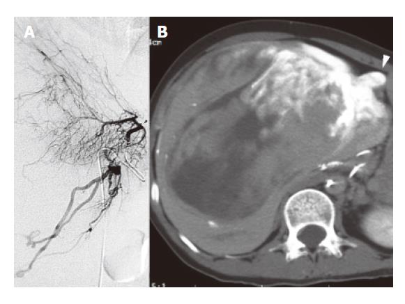
Figure 3 Preoperative angiography.
A: Hepatic arteriography; B: CT during cystic arteriography. Many arteries (i.e., right, middle, left hepatic, and subphrenic arteries fed the tumor (A) (arrow head:the root of cystic artery). However, CT during the selective cystic arteriography revealed stronger enhancement of the tumor than during any other arteriographies (B)(arrow head:gallbladder).
- Citation: Hamada T, Yamagiwa K, Okanami Y, Fujii K, Nakamura I, Mizuno S, Yokoi H, Isaji S, Uemoto S. Primary liposarcoma of gallbladder diagnosed by preoperative imagings: A case report and review of literature. World J Gastroenterol 2006; 12(9): 1472-1475
- URL: https://www.wjgnet.com/1007-9327/full/v12/i9/1472.htm
- DOI: https://dx.doi.org/10.3748/wjg.v12.i9.1472


