INTRODUCTION
Bezoar is adapted from Arabic “bazahr” or “badzehr” which means an antidote or counter-poison due to the fact that till the 19th century, bezoars obtained from sacrificed animals were widely used as antidote[1]. Foreign bodies and bezoars are commonly encountered in children. We describe a child aged 11 years who ingested large amounts of plastic material used for knitting chairs and charpoys. The conglomerate of plastic threads, entrapped food material and other debris, formed a huge mass occupying the whole stomach and even caused ulceration and polyp formation due to irritation of the stomach wall. It is debatable as to whether to call this a bezoar or a foreign body. The appearance, behavior and management of the gastric mass was more like a bezoar than that of a foreign body, and therefore we strongly feel that it should be called a “plastobezoar”.
CASE REPORT
An 11-year-old boy was admitted with complaints of vomiting and abdominal pain of six months duration and swelling over the face and feet for the last one month.
On clinical examination the child was pale with puffiness over the face. Pedal edema was present. Abdominal examination found a firm lump palpable in the epigastrium, which moved with respiration. No crepitus was felt over the lump. There was mild tenderness present over the mass.
The hemoglobin was 5.6 g/dL, total and differential leukocyte counts were normal. The serum urea was 36.4 mmol/L, creatinine 314 μmol/L, and serum albumin was 2 g/dL. Serum globulin, fasting and post-prandial blood glucose, serum amylase, bilirubin and transaminases were normal. Urine examination did not reveal any abnormality. Serum sodium was 135 mmol/L, potassium 2.8 mmol/L and calcium 1.8 mmol/L.
Just after admission, the patient developed typical grandmal epileptic attack, which was managed by intravenous diazepam injection. Intravenous and oral supplements of potassium and calcium were given and the child did not have any further episodes of epilepsy.
The patient was considered to have pre- renal failure due to severe vomiting and dehydration and was managed conservatively with intravenous fluid. Two packs of whole blood were transfused and 100 mL of 20% albumin was given intravenously. Gradually, the serum creatinine and urea became normal. Clinically the puffiness over the face and pedal edema disappeared and the patient started feeling better. Dialysis was not required.
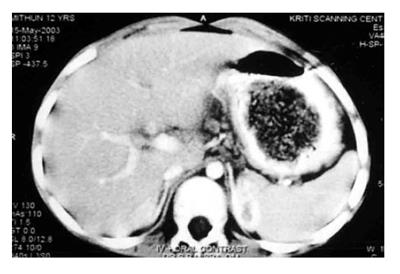
A contrast enhanced CT scan revealed an irregular, large, non-attached mass of varying attenuation with pockets of air, occupying the stomach (Figure 1). A portion of the mass was also encroaching the duodenum. A few polypoidal masses were seen in the antrum of the stomach near the pylorus. There was no post-contrast enhancement of the mass lesion.
Figure 1 Contrast-enhanced CT scan showing a large unattached mass (bezoar) in the stomach with air trapped in it.
There was no post-contrast enhancement of the mass.
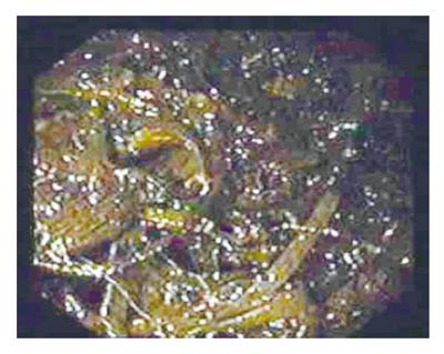
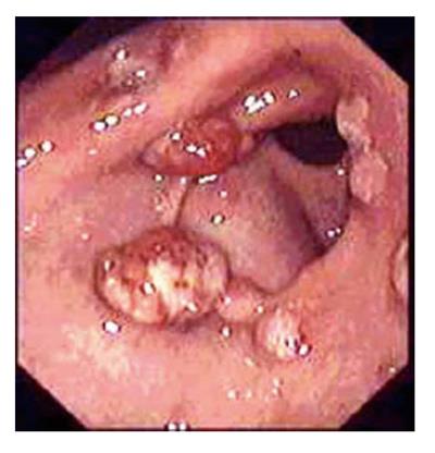
UGI endoscopy revealed a large bezoar occupying nearly the whole stomach (Figure 2). It was black and dark green in color and appeared to be composed of flat plastic thread/grass like material along with entrapped food particles. A small “tail” of the bezoar was also entering the pylorus into the first and second parts of the duodenum. There were several polypoid, masses in the peri-pyloric area of the antrum. Some of the polypoid masses had superficial ulcerations (Figure 3). Ulcerations were also seen at the incisura of the stomach. A few strands of the material forming the bezoar were removed using a rat-toothed forceps. The material was found to be flat plastic threads used in knitting chairs and charpoys. At places, flat cables used for receiving television signals were also found. Attempts to entrap the bezoar in conventional polypectomy snare failed. It could be snared with a large-diameter improvised snare but it could not be delivered through the cardio-esophageal sphincter as one piece due to its size.
Figure 2 Endoscopic appearance of the plastobezoar.
The bezoar was made of hundreds of plastic strands.
Figure 3 Hyperplastic gastric polyps in the pre-pyloric region of the stomach.
On inquiring from the parents of the child, the father said that he had occasionally seen the child peeling off plastic material from chairs and charpoys and eating it for the last three years. It happened when the child was confined to bed (made of plastic) after surgery for an abscess, three years ago, and during this period he started peeling off bits and pieces and strands off the charpoy and swallowing them. Gradually he developed a taste for the plastic material, and even when he recovered he continued to chew and swallow plastic from the charpoys.
The next day, under conscious sedation using injection pentazocine, diazepam and propofol, an overtube was passed into the esophagus over a tightly fitting Savary-Gilliard dilator. Thereafter, the whole bezoar was removed in pieces using rat-toothed forceps, snare and Dormia basket, in an endoscopy session lasting over three hours. Some of the strands from the bezoar measured more than 50 cm. Biopsies were obtained from the polyps in the antrum. The patient was kept nil p.o. overnight. Intravenous pantoprazole was given. A barium meal examination was performed the next day, which did not reveal any abnormality. No obvious delay in the transit time was observed either in the stomach or in the small and large intestines. Psychiatric evaluation did not reveal any abnormality. Histological examination of the biopsy obtained from the polyps in the pre-pyloric region of the stomach revealed the polyps to be hyperplastic in nature. The patient was started on oral pantoprazole and discharged. He is on a regular follow-up about recurrence of the bezoar and disappearance of the hyperplastic polyps.
DISCUSSION
The word bezoar is a word derived from the Arabic “bazahr” or “badzehr” which means an antidote or counter-poison. Till the 19th century, bezoars obtained from sacrificed animals had been widely used as antidote.[1] The origin of most bezoars is thought to be due to delayed gastric emptying as a result of either vagotomy, antral resection, gastroparesis or gastric outlet obstruction[2,3]. However, in a Japanese study, no evidence of delayed gastric emptying was observed[4]. Phytobezoar formation has also been reported to form in an adolescent with achalasia cardia and hypertrophic pyloric stenosis[5] and in one patient each having intestinal pseudo-obstruction[6] and scleroderma[7]. However, our patient did not have any of the above mentioned conditions. Ingestion of large amounts of indigestible solids may also precipitate bezoar formation[2]. The case in discussion had swallowed a large amount of plastic material and since plastic cannot be digested, a bezoar formed in the stomach. Stomach phytobezoars are also known to occur in uremic patients[8] and although in the present case, serum creatinine rose at the time of hospitalization, it was due to protracted vomiting and hypovolemia leading to pre-renal failure and not due to any renal disease per se. The renal functions became normal within a few days by conservative management.
Bezoars are conventionally defined as retained concre-tions of animal or vegetable material in the intestinal tract[1]. Therefore, in the truest sense, our patient did not have a bezoar but will be said to have a foreign body in the stomach. However, the appearance, behavior and management of the gastric mass was exactly like a bezoar and therefore it may aptly be called a “plastobezoar.” Apart from common phytobezoars, trichobezoars and diospyrobezoars, cotton bezoars[9], paper bezoars[10], bezoars due to medication (pharmacobezoars)[11-12], synthetic glue[13] and even iron bezoars[14] have been reported. Considering the fact that even non-organic substances have been responsible for producing bezoars, the case in discussion is a fit case to be considered as a bezoar and not a foreign body.
Our patient had a unique bezoar due to eating disorder in which he was eating plastic material used for knitting chairs and charpoys. This abnormal eating disorder of plastikophagia has been reported earlier too[15]. However, the formation of a giant bezoar and its consequences were unique in this patient.
CT scan in our patient revealed a non-attached gastric mass lesion with air pockets. There was no post-contrast enhancement. This appearance was very similar to that described in a patient with trichobezoar[16,17] and therefore it appears that all bezoars may give a similar appearance on CT scan, irrespective of the contents. The fact that all the bezoars are retained substances of foreign origin gives the typical CT picture of a non-attached mass that does not enhance after intravenous contrast injection. Since the bezoar forms umpteen number of pockets in which air is trapped, this is also visualized on the CT scan.
Surgery is frequently resorted to for the removal of bezoars, especially the larger ones[2,3,18]. However, laparoscopic removal[19,20] and endoscopic extraction of bezoars have also been successful. Use of lithotriptor[21], laser ignited mini-explosives[22], mono- polar diathermy knife[21] and electrohydraulic lithotripsy[23] have been successful in removing the bezoars of varying etiologies. In our patient, surgery was considered to be an option but was abandoned in view of his poor general condition and the presence of renal failure. Since the bezoar was made up of hundreds of thread-like strands of plastic and food debris, we were unable to use the above-mentioned endoscopic techniques. The bezoar could be trapped as one piece in a large self-made snare, which has been used by us earlier for removal of large foreign bodies such as a mango kernel from the esophagus[24], but due to its large size it could not be negotiated across the cardio-esophageal sphincter. Therefore, it had to be removed the hard way in bits and pieces using an overtube, a rat-toothed forceps, a Dormia basket and a polypectomy snare. We did not have a large channel endoscope, which has been used to remove gastric phytobezoars of 500, 700 and 1000 ml successfully in one go by sucking them from the stomach[25].
The hyperplastic gastric polyps (Figure 3), in the pre-pyloric region of the stomach, probably occurred because of chronic irritation of the gastric mucosa by the bezoar. This was also the reason for the ulcerations seen in the stomach. It is logical to think that these polyps may have caused further stasis in the stomach by deforming the pyloric opening and impeding the flow of the gastric contents. It is interesting in this regard to find a couple of case reports where gastric polyposis[26] and hyperplastic gastric polyps[27] were noted to be associated with gastric bezoar formation[4].
S- Editor Wang J L- Editor Ma JY E- Editor Ma WH


