Published online Sep 7, 2006. doi: 10.3748/wjg.v12.i33.5399
Revised: March 28, 2006
Accepted: April 21, 2006
Published online: September 7, 2006
Left-sided periappendiceal abscesses occur in association with two types of congenital anomaly: intestinal malrotation and situs inversus. It is difficult to obtain an accurate preoperative diagnosis of these abscesses due to the abnormal position of the appendix. We present an unusual case of a left-sided periappendiceal abscess in an adult with intestinal malrotation, the diagnosis of which was a challenge.
- Citation: Lee MR, Kim JH, Hwang Y, Kim YK. A left-sided periappendiceal abscess in an adult with intestinal malrotation. World J Gastroenterol 2006; 12(33): 5399-5400
- URL: https://www.wjgnet.com/1007-9327/full/v12/i33/5399.htm
- DOI: https://dx.doi.org/10.3748/wjg.v12.i33.5399
Intestinal malrotation is a congenital anomaly referring to either nonrotation or incomplete rotation of the primitive intestinal loop around the axis of the superior mesenteric artery during fetal development. While most cases of intestinal malrotation present with bilious vomiting in the first month of life[1], rare cases present in adulthood[2]. It is important that physicians be aware of the possibility of this disease when treating adult patients with abdominal pain because diagnosis of intestinal malrotation can be difficult. The present report details an unusual case of a left-sided periappendiceal abscess with intestinal malrotation in an adult who presented with a painful mass in the left lower quadrant.
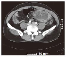
A 43-year-old man with no previous abdominal complaints presented to the emergency room of our hospital with a 2-wk history of a painful mass in the left lower quadrant. Previously, his physician had administered intravenous antibiotics for suspected diverticulitis, but increasing abdominal pain led to his hospital presentation. Physical examination revealed a body temperature of 37.4°C and an 8 cm-sized painful mass in the left lower quadrant. Laboratory tests showed a normal white cell count (4.6 × 10⁹/L) and an elevated C-reactive protein concentration (104 mg/L). Computed tomography (CT) abdominal scanning revealed a solid fluid-containing tumor in the left lower quadrant (Figure 1), suggesting an inflammatory mass with abscess formation. We drained the abscess using percutaneous drainage (PCD) and administered intravenous antibiotics while trying to determine the exact cause of the abscess. One week later, the patient had a soft abdomen with no specific complaint.
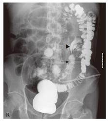
To identify the cause of the abscess, we performed a contrast enema with gastrograffin. This procedure revealed that a fistulous tract of the colon did not exist, and the entire colon was seen in the left half of the abdomen with the cecum in the left lower quadrant (Figure 2). Contrast filling of the terminal ileum was apparent. An irregular contour of the cecum was also observed, with no contrast filling of the appendix. Figure 2 also shows a pig-tail catheter located in the area corresponding to an abscess pocket. With this information, the abdominal CT was again reviewed. Further review indicated a periappendiceal abscess with intestinal malrotation due to the anatomic location of the right-sided small bowel, left-sided large bowel, the abnormal position of the superior mesenteric vessels, and an inflammatory mass in the ileocecal area.
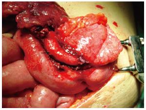
Surgery was performed through a lower midline incision after obtaining informed consent. Surgical findings confirmed the severe inflammatory changes of the appendix and ileocecal region located in the left lower quadrant (Figure 3). An ileocecectomy was performed. Pathology testing indicated gangrenous appendicitis with severe pericecal inflammation. The patient recovered uneventfully after surgery.
Intestinal malrotation is a congenital anomaly referring to either non-rotation or incomplete rotation of the primitive intestinal loop around the axis of the superior mesenteric artery during fetal development. As most complications associated with intestinal malrotation occur in the first month of life[1], this disease may not be foremost in the mind of physicians with exclusively adult patients. However, all such physicians should be familiar with intestinal malrotation owing to its associated diagnostic difficulty and the devastating consequences of failure of recognition[2]. In general, adults with intestinal malrotation present in one of three ways[3]. Some patients present with acute obstructive symptoms and signs of impending abdominal catastrophe. Others present with chronic abdominal complaints that include both pain and intermittent obstruction. Lastly, some present with atypical symptoms common to abdominal diseases unrelated to intestinal malrotation, such as in the present report.
The increasing use of abdominal CT as the first imaging modality in patients with various abdominal complaints means that the importance of identifying malrotation by CT cannot be overemphasized. Malrotation can be diagnosed on CT by the anatomic location of a right-sided small bowel, a left-sided colon, an abnormal relationship of the superior mesenteric vessels, and aplasia of the uncinate process of the pancreas[4]. In the present case, such CT findings indicating malrotation were not immediately recognized due to the low frequency of this disease in the adult population. Malrotation was recognized only after a contrast enema showed that the entire colon was in the left half of the abdomen, with the cecum in the left lower quadrant. Furthermore, left-sided periappendiceal abscess was not suspected initially because there were no clinical characteristics suggesting appendicitis such as preceding vague central pain and there were only a few cases reported in the literature[5].
We debated as to whether correction of the malrotation was indicated in this case. Dietz et al[2] advocated that correction of the malrotation is probably not indicated unless there is evidence of intestinal obstruction. In contrast, Cathcart et al[6] argued that surgical correction is warranted due to the risk of midgut volvulus.
In conclusion, all physicians with exclusively adult patients should be familiar with intestinal malrotation in order to make a timely and correct diagnosis that will lead to prompt and appropriate treatment.
S- Editor Pan BR L- Editor Zhu LH E- Editor Bai SH
| 1. | Rescorla FJ, Shedd FJ, Grosfeld JL, Vane DW, West KW. Anomalies of intestinal rotation in childhood: analysis of 447 cases. Surgery. 1990;108:710-75; discussion 710-75;. [PubMed] |
| 2. | Dietz DW, Walsh RM, Grundfest-Broniatowski S, Lavery IC, Fazio VW, Vogt DP. Intestinal malrotation: a rare but important cause of bowel obstruction in adults. Dis Colon Rectum. 2002;45:1381-1386. [RCA] [PubMed] [DOI] [Full Text] [Cited by in Crossref: 77] [Cited by in RCA: 61] [Article Influence: 2.7] [Reference Citation Analysis (0)] |
| 3. | Kapfer SA, Rappold JF. Intestinal malrotation-not just the pediatric surgeon's problem. J Am Coll Surg. 2004;199:628-635. [RCA] [PubMed] [DOI] [Full Text] [Cited by in Crossref: 97] [Cited by in RCA: 101] [Article Influence: 4.8] [Reference Citation Analysis (0)] |
| 4. | Zissin R, Rathaus V, Oscadchy A, Kots E, Gayer G, Shapiro-Feinberg M. Intestinal malrotation as an incidental finding on CT in adults. Abdom Imaging. 1999;24:550-555. [RCA] [PubMed] [DOI] [Full Text] [Cited by in Crossref: 84] [Cited by in RCA: 77] [Article Influence: 3.0] [Reference Citation Analysis (0)] |
| 5. | Kamiyama T, Fujiyoshi F, Hamada H, Nakajo M, Harada O, Haraguchi Y. Left-sided acute appendicitis with intestinal malrotation. Radiat Med. 2005;23:125-127. [PubMed] |
| 6. | Cathcart RS 3rd, Williamson B, Gregorie HB Jr, Glasow PF. Surgical treatment of midgut nonrotation in the adult patient. Surg Gynecol Obstet. 1981;152:207-210. [PubMed] |


