Published online Apr 21, 2006. doi: 10.3748/wjg.v12.i15.2464
Revised: November 6, 2005
Accepted: November 18, 2005
Published online: April 21, 2006
Although foreign body ingestion is relatively common, toothbrush swallowing is rare. We report a case of a swallowed toothbrush which passed through the ileocecal valve and perforated the proximal transverse colon, then the liver. To our knowledge, this is the first case to be reported.
- Citation: Lee MR, Hwang Y, Kim JH. A case of colohepatic penetration by a swallowed toothbrush. World J Gastroenterol 2006; 12(15): 2464-2465
- URL: https://www.wjgnet.com/1007-9327/full/v12/i15/2464.htm
- DOI: https://dx.doi.org/10.3748/wjg.v12.i15.2464
Toothbrush ingestion is uncommon, but requires prompt medical attention. Although 80% of ingested foreign bo-dies pass spontaneously [1], there are no reports regarding swallowed toothbrushes passing through the pylorus [2]. Here we present an unusual case of a toothbrush swallowing which passed through the ileocecal valve and perfora-ted the proximal transverse colon, then penetrated the liver. To our knowledge, this is the first case to be reported.
A 31 year-old man was admitted to the Surgical Department via the Emergency Room with one week history of right upper abdominal pain. He was diagnosed with schizophrenia 13 years earlier and treated at a local hospital. A physical examination revealed tenderness in the right upper quadrant and a temperature of 37 °C. Laboratory tests showed a white blood cell count in the upper normal range, a slightly elevated C-reactive protein level (23 mg/L) and elevated aspartate aminotransferase/alanine aminotransferase levels (51/99 IU/L).
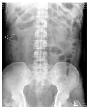
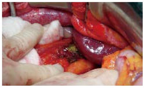
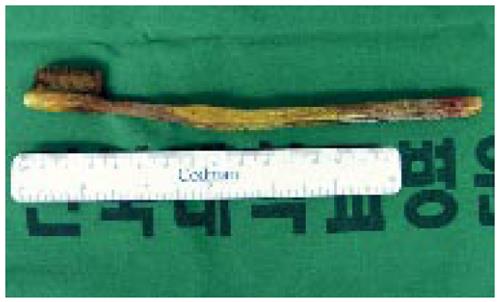
A plain abdominal radiograph showed a characteristic radiographic image of a toothbrush with parallel rows of short metallic radiodensities in the right upper quadrant (Figure 1). At that time, the patient stated he swallowed a toothbrush 1 year earlier. An abdominal computed tomography scan revealed a metallic density in the ascending colon (Figure 2A) and a low density lesion penetrating the lateral section of the liver (Figure 2B). The presumptive diagnosis was colohepatic penetration by a swallowed toothbrush and a laparotomy was performed. No abnormal ascites fluid was observed. Dense adhesions between the proximal transverse colon and the lateral section of the liver were found. When the proximal transverse colon was mobilized, the shaft of a toothbrush was observed penetrating the colon and liver (Figure 3). The toothbrush was removed and the perforated colonic opening was repaired. The extracted toothbrush was 20 cm long (Figure 4). The patient had an uneventful hospital course and was discharged eleven days after surgery.
Ingestion of a foreign body is commonly encountered in the clinic among children, adults with intellectual impairment, psychiatric illness or alcoholism, and dental prosthetic-wearing elderly subjects [1,3]. However, toothbrush swallowing is rare, with only approximately 40 reported cases [2]. It was reported that a toothbrush shows a characteristic radiographic image with parallel rows of short metallic radiodensities due to the metallic plates that hold the bristles in place [4]. Unlike most other foreign bodies, there are no reports of swallowed toothbrushes passing spontaneously [2]. Thus, prompt intervention is required in order to avoid complications such as pressure necrosis causing gastritis, ulceration and perforation [5]. An initial extraction strategy to consider is endoscopy by a skilled technician, and the first successful performance of this procedure has been reported by Ertan et al [6]. If endoscopic removal is not possible and particular complications are not present, a laparoscopic approach may be an alternative to laparotomy [7].
To our knowledge, this is the first report of a swallowed toothbrush passing through the ileocecal valve and penetrating the colon and liver. Similar to the present case, there are reports of toothpicks penetrating the pyloroduodenal region and migrating to the liver [8]. A Medline search indicates that other similar reports involving toothbrushes are found only in the esophagus and stomach. In the present case, it is highly remarkable that a 20 cm toothbrush could pass through the pylorus and duodenal loop.
S- Editor Guo SY L- Editor Wang XL E- Editor Zhang Y
| 1. | Selivanov V, Sheldon GF, Cello JP, Crass RA. Management of foreign body ingestion. Ann Surg. 1984;199:187-191. [RCA] [PubMed] [DOI] [Full Text] [Cited by in Crossref: 141] [Cited by in RCA: 146] [Article Influence: 3.6] [Reference Citation Analysis (0)] |
| 2. | Kirk AD, Bowers BA, Moylan JA, Meyers WC. Toothbrush swallowing. Arch Surg. 1988;123:382-384. [RCA] [PubMed] [DOI] [Full Text] [Cited by in Crossref: 26] [Cited by in RCA: 28] [Article Influence: 0.8] [Reference Citation Analysis (0)] |
| 3. | Velitchkov NG, Grigorov GI, Losanoff JE, Kjossev KT. Ingested foreign bodies of the gastrointestinal tract: retrospective analysis of 542 cases. World J Surg. 1996;20:1001-1005. [RCA] [PubMed] [DOI] [Full Text] [Cited by in Crossref: 303] [Cited by in RCA: 261] [Article Influence: 9.0] [Reference Citation Analysis (0)] |
| 4. | Riddlesberger MM Jr, Cohen HL, Glick PL. The swallowed toothbrush: a radiographic clue of bulimia. Pediatr Radiol. 1991;21:262-264. [RCA] [PubMed] [DOI] [Full Text] [Cited by in Crossref: 20] [Cited by in RCA: 19] [Article Influence: 0.8] [Reference Citation Analysis (0)] |
| 5. | Kaye WH, Klump KL, Frank GK, Strober M. Anorexia and bulimia nervosa. Annu Rev Med. 2000;51:299-313. [RCA] [PubMed] [DOI] [Full Text] [Cited by in Crossref: 104] [Cited by in RCA: 79] [Article Influence: 3.2] [Reference Citation Analysis (0)] |
| 6. | Ertan A, Kedia SM, Agrawal NM, Akdamar K. Endoscopic removal of a toothbrush. Gastrointest Endosc. 1983;29:144-145. [RCA] [PubMed] [DOI] [Full Text] [Cited by in Crossref: 16] [Cited by in RCA: 15] [Article Influence: 0.4] [Reference Citation Analysis (0)] |
| 7. | Wishner JD, Rogers AM. Laparoscopic removal of a swallowed toothbrush. Surg Endosc. 1997;11:472-473. [RCA] [PubMed] [DOI] [Full Text] [Cited by in Crossref: 29] [Cited by in RCA: 32] [Article Influence: 1.1] [Reference Citation Analysis (0)] |
| 8. | Kanazawa S, Ishigaki K, Miyake T, Ishida A, Tabuchi A, Tanemoto K, Tsunoda T. A granulomatous liver abscess which developed after a toothpick penetrated the gastrointestinal tract: report of a case. Surg Today. 2003;33:312-314. [RCA] [PubMed] [DOI] [Full Text] [Cited by in Crossref: 37] [Cited by in RCA: 36] [Article Influence: 1.6] [Reference Citation Analysis (0)] |


