Copyright
©2005 Baishideng Publishing Group Inc.
World J Gastroenterol. Feb 14, 2005; 11(6): 903-907
Published online Feb 14, 2005. doi: 10.3748/wjg.v11.i6.903
Published online Feb 14, 2005. doi: 10.3748/wjg.v11.i6.903
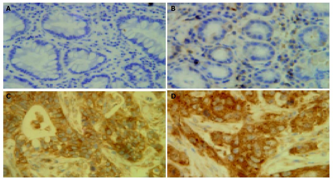
Figure 1 Comparison of nuclear staining rates statistically higher in tumor cells with those in adjacent and normal epithelial cells after staining of RelA.
A: Normal epithelial cells with no nuclei staining; B: Adjacent epithelial cells with no nuclei staining; C: Cancer tissues with strong nuclei staining; D: Cancer metastasis to the liver with specially enhanced nuclei staining (×400).
- Citation: Cao HJ, Fang Y, Zhang X, Chen WJ, Zhou WP, Wang H, Wang LB, Wu JM. Tumor metastasis and the reciprocal regulation of heparanase gene expression by nuclear factor kappa B in human gastric carcinoma tissue. World J Gastroenterol 2005; 11(6): 903-907
- URL: https://www.wjgnet.com/1007-9327/full/v11/i6/903.htm
- DOI: https://dx.doi.org/10.3748/wjg.v11.i6.903


