Published online Dec 28, 2005. doi: 10.3748/wjg.v11.i48.7694
Revised: June 13, 2005
Accepted: June 18, 2005
Published online: December 28, 2005
Minimally invasive surgery has revolutionized the treatment of gastrointestinal tumors. Submucosal tumors of the stomach can be resected using laparoscopic techniques. We report here a case of ectopic pancreas tissue in the gastric wall that was removed using robotic-assisted laparoscopic resection. The patient was a 15-year-old female who presented with abdominal discomfort and tarry stools. Laboratory analysis showed iron deficiency anemia. Preoperative endoscopy revealed a submucosal lesion in the posterior wall of the gastric high body. Intraoperative upper endoscopy clearly located the lesion. A robotic-assisted laparoscopic wedge resection of the putative gastric submucosal tumor was performed. The pathology results showed an ectopic pancreas. The patient had an uneventful recovery and we believe that this is a valid treatment option for this benign condition.
- Citation: Hsu SD, Wu HS, Kuo CL, Lee YT. Robotic-assisted laparoscopic resection of ectopic pancreas in the posterior wall of gastric high body: Case report and review of the literature. World J Gastroenterol 2005; 11(48): 7694-7696
- URL: https://www.wjgnet.com/1007-9327/full/v11/i48/7694.htm
- DOI: https://dx.doi.org/10.3748/wjg.v11.i48.7694
Ectopic pancreas is relatively rare and is definite as abnormally situated pancreatic tissue has no contact with the normal pancreas, and has its own ductal system and blood supply[1]. It is a rare entity and is usually an incidental finding in clinical practice. Most patients with an ectopic pancreas are asymptomatic, and if present, symptoms are nonspecific and depend upon the site of the lesion and different complications are encountered. Heterotopic pancreatic tissue has been found in several abdominal and intrathoracic locations, most frequently in the stomach (25-60%) or duodenum (25-35%)[2]. We report here a successful robotic-assisted laparoscopic wedge resection of a putative gastric submucosal tumor in a 15-year-old female.
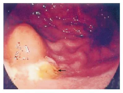
A 15-year-old adolescent girl presented with intermittent epigastric pain with tarry stools for about 10 mo. She did not have any history of gastrointestinal cancer. The familial and personal history was nothing special. Laboratory findings showed iron deficiency anemia that prompted further gastrointestinal evaluation. The results of abdominal sonography and colonoscopy were negative. Panendoscopy revealed a submucosal lesion in the posterior wall of the gastric high body (Figure 1). The submucosal lesion measured about 1.5 cm in diameter. Pathological evaluation of a biopsy sample revealed chronic gastritis and mucosal hyperplasia. Biopsies collected with endoscopic techniques often do not provide the representative histologic sample needed for further therapeutic decisions. Because a malignant etiology could not be ruled out, a laparoscopic-endoscopic approach was considered to be appropriate for a curative and definitive diagnosis and minimally invasive for the resection of a localized gastric submucosal tumor.
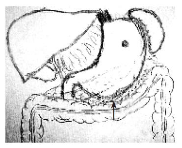
The initial trocar was inserted at the umbilicus, using the Hasson technique. A pneumoperitoneum was created with carbon dioxide. In total, four trocars were inserted in the upper abdomen. The abdominal cavity was fully explored, during which robotic-assisted laparoscopic procedures were performed using the Zeus robot system. The greater omentum was detached from the greater curvature of the stomach with a harmonic scalpel, and the lesser sac was entered (Figure 2).
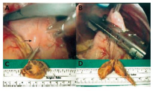
A clearly localized tumor mass was identified with the assistance of intraoperative upper-tract endoscopy. A robotic-assisted laparoscopic wedge resection of the gastric high body was performed using the Endo-GIA roticulator (Figure 3). The resection margins were clear and the specimen was sent for pathological analysis. A layer of interrupted silk sutures was placed in the serosa surface to ensure the integrity of the staple line. A nasogastric tube was inserted into the stomach and a drain tube was placed near the staple line. The abdominal cavity was deflated and the trocar sites were closed.
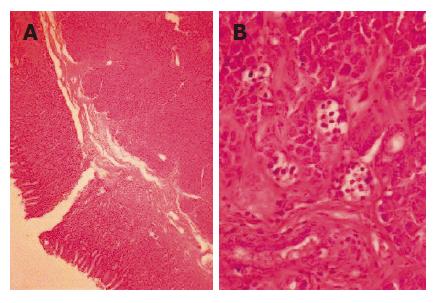
The final pathology report revealed that the resected specimen was an ectopic pancreas (Figure 4). The patient was discharged on postoperative d 6, with no complications and normal gastrointestinal motility. She had an uneventful recovery.
Ectopic pancreas is a rare entity and is usually an incidental finding in clinical practice. Most patients with an ectopic pancreas are asymptomatic, and if present, symptoms are nonspecific and depend on the site of the lesion and the different complications are encountered[3-5]. About 75% of all pancreatic rests are located in the stomach, duodenum, or jejunum[6]. However, they have also been found in the ileum, Meckel’s diverticulum, gall bladder, common bile duct, splenic hilum, umbilicus, lung, and in perigastric and periduodenal tissues[7]. In autopsy series, the frequency of ectopic pancreas is between 1% and 2%. The rate of recognition at the time of laparotomy is 0.2%.
A preoperative diagnosis of ectopic pancreas in the gastric wall is not easy. Although a radiological diagnosis of gastric ectopic pancreatic tissue is difficult, double-contrast barium meal may show a characteristic focally raised mucosal area with associated superficial ulceration. The role of endoscopic biopsies in identifying ectopic pancreas remains questionable because normal gastric mucosa covers the lesion. Recently, endoscopic ultrasound combined with fine-needle aspiration cytology has been reported to facilitate a definitive diagnosis by histological examination[8]. Surgical excision, either endoscopically or laparoscopically, provides symptomatic relief and is recommended if the diagnosis remains uncertain. Laparoscopic wedge resection of a presumed gastric submucosal tumor appears to have been a suitable treatment for our patient. This approach is more advantageous over a laparotomy because recovery is easier and morbidity is less.
With the advent of laparoscopy at the end of the 1980s, surgery has entered the computer age[9]. More recently, robotic-assisted laparoscopy has joined the general surgeon’s armory to address some of the shortcomings of laparoscopic surgery. Magnified and computer-enhanced video images provide surgeons with much better access to and visualization of the abdomen. The main advantages of robot-assisted laparoscopic surgery are the availability of three-dimensional visibility and easier instrument manipulation compared to standard laparoscopy. Initially, robotic-assisted laparoscopic cholecystectomy was deemed safe, and has been proved to be safe in foregut procedures.
In conclusion, ectopic pancreas in the posterior wall of gastric high body can be resected by robotic-assisted laparotomy. This procedure is minimally invasive for such benign lesions.
Science Editors Wang XL and Guo SY Language Editor Elsevier HK
| 1. | Guillou L, Nordback P, Gerber C, Schneider RP. Ductal adenocarcinoma arising in a heterotopic pancreas situated in a hiatal hernia. Arch Pathol Lab Med. 1994;118:568-571. [PubMed] |
| 2. | Moen J, Mack E. Small-bowel obstruction caused by heterotopic pancreas in an adult. Am Surg. 1989;55:503-504. [PubMed] |
| 3. | Mulholland MW, Simeone DM. Pancreas: Anatomy and structural anomalies: Congenital anomalies: Heterotopic pancreas. Philadelphia: Lippincott Williams & Wilkins 1999; 2115-2119. |
| 4. | Abrahams JI. Heterotopic pancreas simulating peptic ulceration. Arch Surg. 1966;93:589-592. [RCA] [PubMed] [DOI] [Full Text] [Cited by in Crossref: 9] [Cited by in RCA: 8] [Article Influence: 0.1] [Reference Citation Analysis (0)] |
| 5. | Armstrong CP, King PM, Dixon JM, Macleod IB. The clinical significance of heterotopic pancreas in the gastrointestinal tract. Br J Surg. 1981;68:384-387. [RCA] [PubMed] [DOI] [Full Text] [Cited by in Crossref: 130] [Cited by in RCA: 130] [Article Influence: 3.0] [Reference Citation Analysis (0)] |
| 6. | Grendell JH, Ermak TH. Anatomy, Histology, Embriology, and Developmental Anomalies of the Pancreas. In: Sleisenger & Fordtran's Gastrointestinal and Liver Disease, Philadelphia: WBSaunders 1998; 761-771. |
| 7. | Kopelman HR. The pancreas: Congenital anomalies. Pediatric Gastrointestinal Disease. St. Lous: Mosb 1996; 1426-1427. |
| 8. | Riyaz A, Cohen H. Ectopic pancreas presenting as a submucosal gastric antral tumor that was cystic on EUS. Gastrointest Endosc. 2001;53:675-677. [RCA] [PubMed] [DOI] [Full Text] [Cited by in Crossref: 10] [Cited by in RCA: 16] [Article Influence: 0.7] [Reference Citation Analysis (0)] |
| 9. | Chapman WH, Albrecht RJ, Kim VB, Young JA, Chitwood WR. Computer-assisted laparoscopic splenectomy with the da Vinci surgical robot. J Laparoendosc Adv Surg Tech A. 2002;12:155-159. [RCA] [PubMed] [DOI] [Full Text] [Cited by in Crossref: 37] [Cited by in RCA: 27] [Article Influence: 1.2] [Reference Citation Analysis (0)] |


