Copyright
©2005 Baishideng Publishing Group Inc.
World J Gastroenterol. Nov 28, 2005; 11(44): 6954-6959
Published online Nov 28, 2005. doi: 10.3748/wjg.v11.i44.6954
Published online Nov 28, 2005. doi: 10.3748/wjg.v11.i44.6954
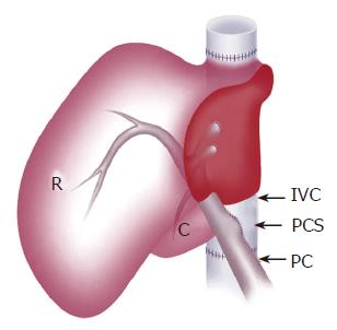
Figure 2 Schematic view of the liver graft with PCS placed by side-to-side anastomosis of PV and IVC.
R: right lateral lobe; C: caudate lobe; PV: portal vein; IVC: inferior vena cava; PCS: portocaval shunt.
- Citation: Wang HS, Ohkohchi N, Enomoto Y, Usuda M, Miyagi S, Asakura T, Masuoka H, Aiso T, Fukushima K, Narita T, Yamaya H, Nakamura A, Sekiguchi S, Kawagishi N, Sato A, Satomi S. Excessive portal flow causes graft failure in extremely small-for-size liver transplantation in pigs. World J Gastroenterol 2005; 11(44): 6954-6959
- URL: https://www.wjgnet.com/1007-9327/full/v11/i44/6954.htm
- DOI: https://dx.doi.org/10.3748/wjg.v11.i44.6954


