Published online Sep 14, 2005. doi: 10.3748/wjg.v11.i34.5336
Revised: April 1, 2005
Accepted: April 2, 2005
Published online: September 14, 2005
AIM: To investigate the role of SPECT/CT in the diagnosis of hepatic hemangiomas whose anatomical positions are not ideal, situated adjacent to the heart, the inferior cava, hepatic vessels or abdominal aorta, etc.
METHODS: The hepatic perfusion, blood pool, and fusion imaging were carried out using SPECT/CT in 54 patients, who were suspected for hepatic hemangiomas. When the anatomical positions were not ideal, the diagnosis was difficult by SPECT only. So the information of computed tomography (CT) was applied to help in diagnosing. The results were recorded as hemangiomas or not.
RESULTS: Of the 54 patients, 31 patients were diagnosed as suffering from hepatic hemangiomas. The anatomical positions of eight patients’ hepatic hemangiomas (25.81%) were not ideal. Among these lesions of the eight patients, three patients’ hepatic lesions were located near to the abdominal aorta, one to the heart, and four to the inferior cava. In addition, six abnormal radioactivity accumulation regions, adjacent to the heart and inferior cava, with the help of CT, were confirmed to be the imaging of inferior cava other than hepatic hemangiomas.
CONCLUSION: When the anatomical positions of hepatic hemangiomas are not good enough for diagnosis, the fusion imaging of SPECT/CT is a simple and efficient method for differential diagnosis.
- Citation: Zheng JG, Yao ZM, Shu CY, Zhang Y, Zhang X. Role of SPECT/CT in diagnosis of hepatic hemangiomas. World J Gastroenterol 2005; 11(34): 5336-5341
- URL: https://www.wjgnet.com/1007-9327/full/v11/i34/5336.htm
- DOI: https://dx.doi.org/10.3748/wjg.v11.i34.5336
The diagnosis of a number of atypical hepatic hemangiomas in cross-sectional images is also very difficult[1]. Moreover, when the tumor is present in staging or in the follow-up of oncological patients, the differentiation of hepatic hemangioma from hepatic metastatistic diseases is a common clinical question. Contrast-enhanced magnetic resonance imaging (MRI) and multiple helical computed tomography (CT) is also limited to distinguish focal hepatic lesions in some ways[2]. Contrast-enhanced ultrasonography (US) is helpful to confirm the characterization of the focal liver disease. However, the diagnosis is associated with the experience of the surgeon. As regards single-photon emission computed tomography (SPECT), when the anatomical positions of hepatic hemangiomas were not ideal, for example, close to the heart, inferior cava or main hepatic vessels, etc., it may cause some difficulty for diagnosis, because the radioactivity of the 99mTc-labeled RBC is centralized in the heart, inferior cava, abdominal aorta and main hepatic vessels, and the radioactivity of blood pool is existing from the start. Thus, it is difficult to detect hepatic hemangioma, which is close to the above-mentioned structure, especially tiny ones.
A method, SPECT/CT, which can localize the hepatic lesions accurately will be helpful to diagnose this kind of hepatic hemangioma. The imaging of 99mTc-labeled red blood cell scintigraphy through SPECT is non-invasive and highly specific, able to avoid invasive method or biopsy. Meanwhile, when the anatomical position is not ideal, close to heart, inferior cava, main hepatic vessels, abdominal aorta, upside of right kidney, the diagnosis accuracy is improved owing to the locating function of the CT.
Fifty-four patients who were suspected for hepatic hemangiomas found by US, CT, and or MRI, and were examined by SPECT/CT from April 26, 2001 to July 15, 2004 in Beijing Hospital. Their average age was 49.15 ± 13.78 years. Among them, 31 were men with an average age of 50.38 ± 15.36 years; 23 were women with an average age of 47.36 ± 11.22 years. The oldest was 91 years old and the youngest was 27 years old.
First, the patients were required to take 400 mg of perchlorate potassium. Thirty minutes later, 10 mg of pyrophosphate dissolved in 2 mL of water for injection was administered into one patient. Thirty minutes later again, 740 MBq of Na99mTcO4 was also administered intravenously by bolus injection. The imaging was obtained using the SPECT/CT (Hawkeye, General Electronic Medical System).
The patients were laid on the table and the detector was placed at front-back imaging position or back-front one. The field of vision included parts of heart, abdominal aorta, liver, spleen, and kidney. The SPECT was equipped with low-energy parallel hole general collimator or high-resolution collimator. The energy peak was 140 keV, the width of the energy window was from 15% to 20%, and the matrix 128×128. When Na99mTcO4 was injected through elbow vein by bolus, the acquisition was carried out immediately at the speed of 2 s/frame, totally 30 frames.
The static imaging was obtained at 5, 10, 15, and 20 min after intravenous injection. The delayed imaging was obtained from 1.5 to 2 h after injection, and 4-6 h if necessary.
Planar imaging: The patients were laid on the table. The detector was placed at the front-back and back-front position, or in the position where hepatic lesions were able to be shown clearly. The detectors were equipped with low-energy parallel hole general collimator or high-resolution collimator. The energy peak was 140 keV, the width of the window 20%, and the matrix 256×256. The count was set up 500 K/frame in advance.
The hepatic blood pool cross-sectional imaging was acquired and together with CT scanning after 40 min of bolus intravenous injection. The X-ray transmission scanning, which needed 0.8 min, was obtained firstly with the matrix of 384×800 to identify the liver position. Then X-CT acquisition, which needed 10.8 min, was followed with matrix of 256×256, zoom of 1.105, 10 mm per layer, totally 40 cross-sections. After transmission process, the detector automatically came back to the start position. Then SPECT acquisition over 360° was performed with the matrix of 128×128, zoom of 1.28, 20 s/frame, and 6°/frame. The technique of automatic human body contour was applied to making detector get closer to the patient’s body.
The SPECT images were reconstructed from the raw data by filtered back projection using a HANN filter (filter parameter: 0.85). The CT data were reconstructed using nuclear medicine workstation (eNTEGRA, General Electronic Medical System). Then the matching emission and transmission were fused to form the fusion images.
The standard of diagnosis is that an increase of blood pool activity within a liver lesion is typical of hemangioma. And in the CT scanning, the density of the lesion is low and even. The images were read by experienced physicians, who have worked in the department of nuclear medicine for at least 5 years. The results were recorded as hepatic hemangioma or not.
Of the 54 patients, 31 (57.41%) were diagnosed as hepatic hemangioma patients by SPECT/CT and were not taken for further examination. The diameter of hepatic lesions was from 1 to 9 cm. Twenty-three patients (42.59%) were excluded from the hepatic hemangioma, because their hepatic lesions did not meet the characterization of the hepatic hemangioma. Among them, 9 patients lost contact with us, the 13 patients’ were diagnosed as hepatic cancer, and one as hepatic cyst.
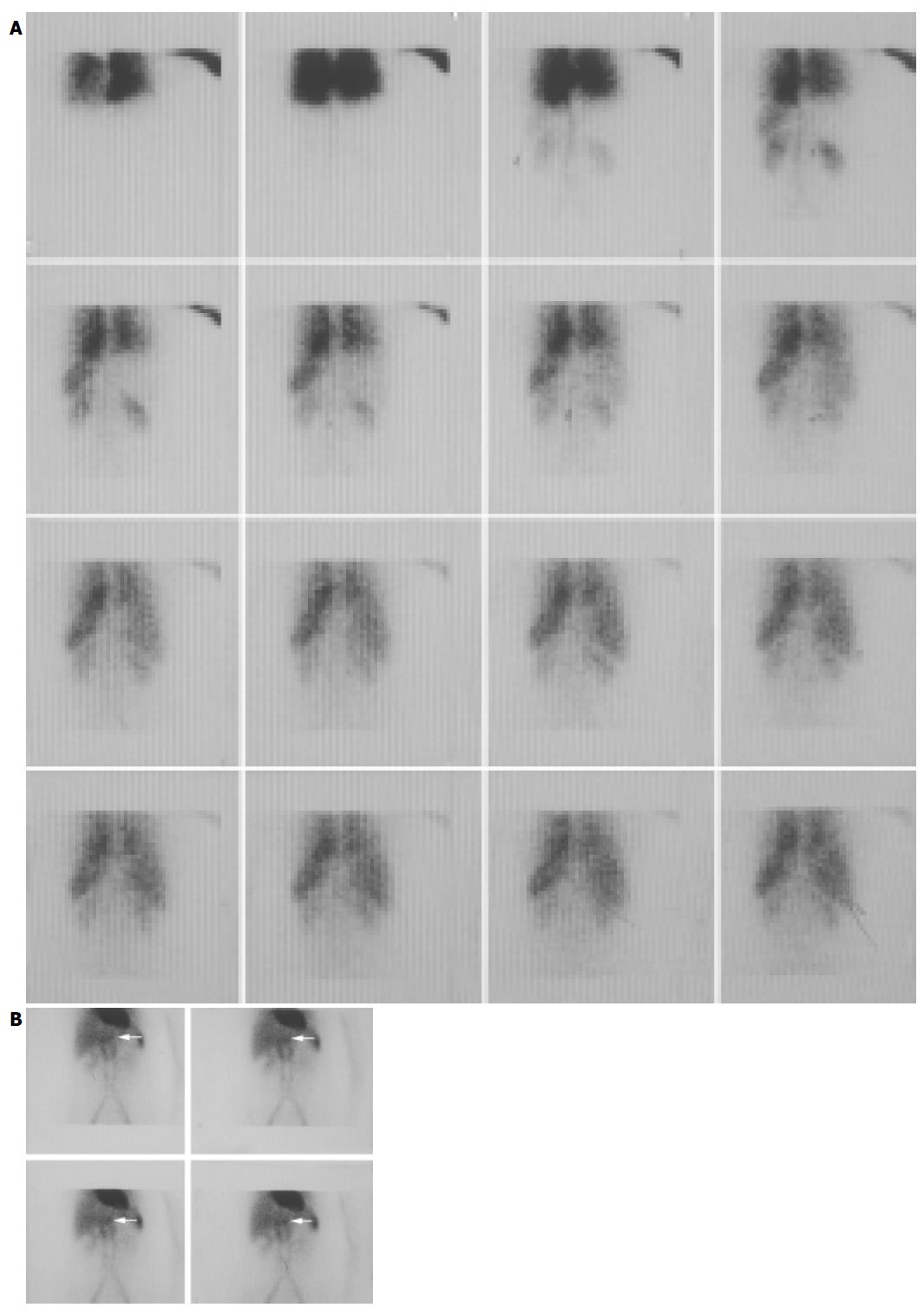
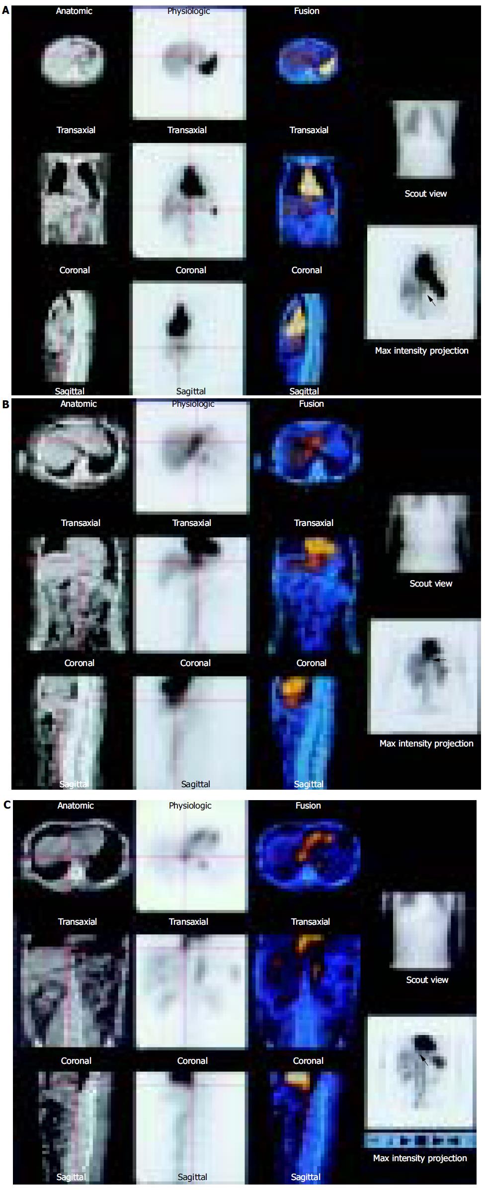
Of the 31 patients, the anatomical positions of the hepatic hemangiomas in 8 patients (25.81%) were not ideal. Among them, the hepatic lesions of three patients’ were close to abdominal aorta (Figures 1A and B and 2A), that of four patients to inferior cava, that of one patient to heart (Figure 2B). Under these conditions, it was difficult to diagnose by only using SPECT. In SPECT/CT fusion imaging, CT was able to locate the position of focal radioactivity accumulation region, and at last diagnosed it as hepatic hemangioma.
In the research, there are six round-like regions with high radioactivity, close to the heart and inferior cava in six patients on the brim of the liver. The CT showed that they were in the inferior cava. Finally, hepatic hemangioma was excluded (Figure 2C).
As a benign tumor, the hepatic hemangioma most frequently appeared in the liver. The incidence of the disease is about from 2% to 7% of the liver lesions. The hepatic hemangioma is classified as capillary and cavernous hepatic hemangioma. The former is not common, and able to change to the other. Thus, the majority is cavernous hepatic hemangioma.
As a non-invasive and highly specific method for diagnosing hepatic hemangioma, the 99mTc-labeled RBC scintigraphy can avoid invasive imaging or biopsy. And thanks to the application of SPECT, the sensitivity of the method is improved obviously. However, when the diameter of hepatic lesion is less than 1.3 cm, the sensitivity of SPECT is quite low. When the diameter is less than 1 cm, false negative appeared. And when the larger hepatic lesions (diameter more than 2.5 cm) with the bad anatomical positions, close to the heart, inferior cava or main hepatic vessels, etc., the diagnosis is difficult in a way, especially tiny hepatic hemangiomas. As reported by Birnbaum et al[3] the hepatic lesions of 18 patients, whose hepatic hemangiomas were adjacent to the above-mentioned positions, 6 of the patients were not able to find them out. As reported by Schillaci et al[4] 24 patients were sceptical of suffering from hepatic hemangiomas. Of them, six patients’ anatomical position were not ideal; three patients’ were close to main hepatic vessel, and two close to inferior cava, one close to heart. When SPECT/CT was carried out, with the help of CT, four were diagnosed as hepatic hemangioma, two were excluded from the hepatic hemangioma. In this research, about 25.81% patients’ anatomical position was not ideal. When SPECT/CT was applied, with the help of CT, finally hepatic hemangioma was diagnosed.
The functional information from the SPECT combining with the anatomical information from the CT is able to solve the problem that anatomical position of hepatic hemangioma is not ideal. CT can accurately locate the lesion of radioactivity concentration region. Several years ago, a new apparatus was discovered. It combined the rotatable double-detectors camera γ with low-dose X-rays tube. The camera γ/CT machine would obtain SPECT and CT images respectively. The projection of X-ray was helpful to find the location of the radioactivity concentration focus. This was SPECT/CT fusion imaging. The SPECT/CT was used to diagnose hepatic hemangioma with the following advantages.
A. CT can accurately locate the position of radioactivity accumulating focus theoretically. However, the quality of the present CT in SPECT/CT is not very good. Under some conditions, it is very difficult to indicate the position accurately. But it is believed when multiple helical CT is used in fusion imaging in the future, the position of the hepatic lesions will be located accurately. Moreover, it may reach the function of diagnosis CT but not only in locating the position of the disease.
B. When hepatic lesions are detected by CT scanning, the hepatic colloid imaging may not be necessary. The colloid imaging is mainly used to find out the lesion of the liver. But CT is more superior to the hepatic colloid imaging in this respect. Its sensitivity is much higher than the hepatic colloid imaging. Therefore, when hepatic lesions are detected by CT scanning, the hepatic colloid imaging is not necessary.
C. Fusion imaging can be attenuation-corrected by X-ray. Therefore, the quality of the images would be improved. Because of the high quantity of information of X-ray,
especially when the injected dose of 99mTc-labeled RBC is comparatively low and the patient is fat, the quality of the images will be improved obviously.
D. Improving the accuracy of diagnosis. Typical characterization of hepatic hemangioma is that its density is even and low by general CT scanning. The worth of common CT is limited. Usually it indicates the position of hepatic lesions, which is radioactive accumulation region. It provides anatomical information for hepatic hemangioma. SPECT can provide the functional information for pathological changes. Functional information coordinates with anatomical information, in this way the accuracy of diagnosis is improved.
Furthermore, it is worth paying attention to that, when inferior cava passes by the liver, and enters right heart, a great majority can form a round-like radioactive accumulation region on the brim of the liver (Figure 2C). Under the condition, with the help of CT, the radioactive concentration region may be seen under the heart, link with the inferior cava and out of the liver. If it is, then hepatic hemangioma can be excluded. If it is not and the, the focus is in the liver, the diagnosis of hepatic hemangioma could be made.
Science Editor Guo SY Language Editor Elsevier HK
| 1. | Jang HJ, Kim TK, Lim HK, Park SJ, Sim JS, Kim HY, Lee JH. Hepatic hemangioma: atypical appearances on CT, MR imaging, and sonography. AJR Am J Roentgenol. 2003;180:135-141. [RCA] [PubMed] [DOI] [Full Text] [Cited by in Crossref: 101] [Cited by in RCA: 77] [Article Influence: 3.5] [Reference Citation Analysis (0)] |
| 2. | Zheng WW, Zhou KR, Chen ZW, Shen JZ, Chen CZ, Zhang SJ. Characterization of focal hepatic lesions with SPIO-enhanced MRI. World J Gastroenterol. 2002;8:82-86. [PubMed] |
| 3. | Birnbaum BA, Weinreb JC, Megibow AJ, Sanger JJ, Lubat E, Kanamuller H, Noz ME, Bosniak MA. Definitive diagnosis of hepatic hemangiomas: MR imaging versus Tc-99m-labeled red blood cell SPECT. Radiology. 1990;176:95-101. [RCA] [PubMed] [DOI] [Full Text] [Cited by in Crossref: 88] [Cited by in RCA: 87] [Article Influence: 2.5] [Reference Citation Analysis (0)] |
| 4. | Schillaci O, Danieli R, Manni C, Capoccetti F, Simonetti G. Technetium-99m-labelled red blood cell imaging in the diagnosis of hepatic haemangiomas: the role of SPECT/CT with a hybrid camera. Eur J Nucl Med Mol Imaging. 2004;31:1011-1015. [RCA] [PubMed] [DOI] [Full Text] [Cited by in Crossref: 35] [Cited by in RCA: 21] [Article Influence: 1.0] [Reference Citation Analysis (0)] |


