Published online Aug 15, 2004. doi: 10.3748/wjg.v10.i16.2452
Revised: March 26, 2004
Accepted: April 9, 2004
Published online: August 15, 2004
Granular cell tumor (GCT) is uncommon in the colon and rectum. Here we report a case of GCT in the transverse colon. A 48-year-old male patient underwent a screening colonoscopy. A yellowish sessile lesion, about 4 mm in diameter, was found in the transverse colon. An endoscopic snare resection was performed without complication. Histological examination revealed the tumor consisted of plump neoplastic cells with abundant granular eosinophilic cytoplasm containing acidophilic periodic acid Schiff-positive, diastase-resistant granules. Immunohistochemical analysis showed the tumor cells expressed S-100 protein and neuron-specific enolase. Thus, the resected tumor was diagnosed as a GCT. Since GCTs are usually benign, endoscopic resection constitutes an easy and safe treatment. Colonoscopists should consider the possibility of GCT in the differential diagnosis of submucosal tumors of the colon.
- Citation: Sohn DK, Choi HS, Chang YS, Huh JM, Kim DH, Kim DY, Kim YH, Chang HJ, Jung KH, Jeong SY. Granular cell tumor of colon: Report of a case and review of literature. World J Gastroenterol 2004; 10(16): 2452-2454
- URL: https://www.wjgnet.com/1007-9327/full/v10/i16/2452.htm
- DOI: https://dx.doi.org/10.3748/wjg.v10.i16.2452
Granular cell tumor (GCT) is relatively rare soft tissue tumor that can be located anywhere in the body. It commonly occurs in oral cavities and subcutaneous tissues, but is uncommon in the colon and rectum[1,2]. In the gastrointestinal tract, the most common site for GCT is the esophagus, followed by the duodenum, anus and stomach[3,4]. This usually benign tumor appears as a submucosal nodule, measuring less than 2 cm in diameter, and is often found incidentally during colorectal examinations[2-6]. Here we report a case of a 48-year-old man diagnosed with a GCT arising in the transverse colon and treated by endoscopic resection.
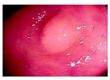
A 48-year-old man was admitted for a screening colonoscopy. He had been healthy without specific complaints, family or past medical history. At the time of colonoscopy, a yellowish, hemispheric nodule 4 mm in diameter, was found in the transverse colon. It was a firm nodule covered by intact mucosa (Figure 1). The patient underwent an endoscopic snare resection, was observed for 30 min, and then discharged. There was no immediate or delayed complication.
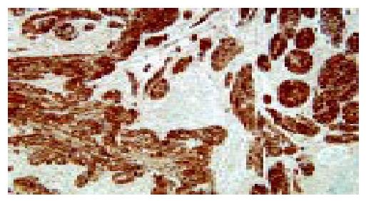
Histological examination of the resected tissue revealed a submucosal tumor composed of solid masses of plump histiocyte-like tumor cells with abundant granular eosinophilic cytoplasm containing acidophilic periodic acid Schiff (PAS)-positive, diastase-resistant granules (Figure 2). Immunohistochemical analysis showed the tumor cells expressed S-100 protein and neuron-specific enolase (NSE) (Figure 3), but were negative for desmin and cytokeratin. The resected tumor was diagnosed as a GCT occurring in the transverse colon.
GCT, first described by Abrikossoff in 1926[7], may arise virtually anywhere in the body, but was seldomly found in the gastrointestinal tract[1,2]. In most cases, gastrointestinal GCTs were found incidentally during endoscopy, and appeared as small, round submucosal nodules covered by normal mucosa[1-5,8-10]. GCTs have also been detected in the muscle layer of the gastrointestinal tract and in subserosal areas, although such findings were uncommon[2,4,11].
Endo et al[10] reported 33 cases of colorectal GCT in Japan, and Rossi et al[8] reported 55 patients diagnosed with GCTs of the colon. To date, 7 cases of colonic GCT have been reported in Korean literature (Table 1). This excludes two cases of perianal GCT which may have arisen from perianal skin rather than anal mucosa. Except for one rectal case, all 6 colonic GCTs reported in Korea were located in the proximal colon - ascending colon, transverse colon and cecum including the appendix. The male-to-female ratio was 1:1.3 and the mean age was 41.0 ± 6.5 years (range 31-49 years). Bowel resection was the treatment administered for the 2 cases diagnosed before 1985, while endoscopic removal of the tumor was performed in the later cases. Advances in endoscopic diagnosis and resection probably led to the alteration in treatment approach for the latter cases.
| N | Year | Author | Sex/Age | Location | Size (cm) | Treatment |
| 1 | 1982 | Kim et al[12] | F/44 | Cecum | 1.5 × 1.5 | Surgery |
| 2 | 1983 | Lee et al[13] | F/31 | Cecum | 1.0 | Surgery |
| 3 | 1991 | Choi et al[14] | F/39 | A-colon | 0.9 × 0.8 | Polypectomy |
| 4 | 2000 | Kim et al [15] | M/40 | Appendix | 0.7 | Polypectomy |
| 5 | 2003 | Lee et al[16] | F/36 | A-colon | 1.5 × 0.6 | Polypectomy |
| 6 | 2003 | Kim et al[17] | M/49 | Rectum | 0.7 | Polypectomy |
| 7 | The present case | M/48 | T-colon | 0.4 × 0.3 | Polypectomy | |
GCT could seldomly be diagnosed based on macroscopic and endoscopic examination due to both its small size and its shape resembling a diminutive polyp[1]. Recently, endoscopic ultrasound has been frequently used for determining the depth of tumor invasion in the gastrointestinal wall, and is also useful for evaluating gastrointestinal submucosal tumors[9,10,18]. However, in the present case, it was difficult to suspect GCT because the endoscopic features of the tumor resembled those of a small sessile polyp. We performed one-stage endoscopic snare polypectomy during screening colonoscopy.
The final diagnosis of GCT depends on pathological findings. The histological markers for GCT are: (1) plump histiocyte-like, bland-looking neoplastic cells with abundant granular eosinophilic cytoplasm containing acidophilic, PAS-positive, diastase-resistant granules; (2) small, uniform nuclei, in which mitotic figures are absent; (3) neural markers, including S-100 protein or NSE expressed uniformly[10,19-22]. In the present patient, the histological findings on the resected specimen were typical of a GCT.
Although GCTs are usually benign, some malignant GCT cases have been reported. Malignancy has been found to correlate with tumor size, more than 60% of metastatic GCTs were larger than 4 cm in diameter[6,11,23]. However, in most colonic GCTs, the tumor size was less than 2 cm and the tumor was well separated from the muscularis propia. Since this tumor is considered to be usually benign, endoscopic removal has been the most appropriate choice of treatment for colonic GCT[2,8,10,24].
In conclusion, we report a case of GCT in the transverse colon. The tumor was removed by endoscopic resection and the patient was discharged without complication. GCTs of the colon can be found incidentally during colonoscopy, and endoscopic removal of the tumor is the safest and most feasible treatment. Colonoscopists should consider the possibility of GCT in the differential diagnosis of submucosal tumors of the colon.
Edited by Wang XL Proofread by Chen WW and Xu FM
| 1. | Lack EE, Worsham GF, Callihan MD, Crawford BE, Klappenbach S, Rowden G, Chun B. Granular cell tumor: a clinicopathologic study of 110 patients. J Surg Oncol. 1980;13:301-316. [RCA] [PubMed] [DOI] [Full Text] [Cited by in Crossref: 415] [Cited by in RCA: 363] [Article Influence: 8.1] [Reference Citation Analysis (0)] |
| 2. | Yasuda I, Tomita E, Nagura K, Nishigaki Y, Yamada O, Kachi H. Endoscopic removal of granular cell tumors. Gastrointest Endosc. 1995;41:163-167. [RCA] [PubMed] [DOI] [Full Text] [Cited by in Crossref: 51] [Cited by in RCA: 43] [Article Influence: 1.4] [Reference Citation Analysis (0)] |
| 3. | Melo CR, Melo IS, Schmitt FC, Fagundes R, Amendola D. Multicentric granular cell tumor of the colon: report of a patient with 52 tumors. Am J Gastroenterol. 1993;88:1785-1787. [PubMed] |
| 4. | Yamaguchi K, Maeda S, Kitamura K. Granular cell tumor of the stomach coincident with two early gastric carcinomas. Am J Gastroenterol. 1989;84:656-659. [PubMed] |
| 5. | Kulaylat MN, King B. Granular cell tumor of the colon. Dis Colon Rectum. 1996;39:711. [RCA] [PubMed] [DOI] [Full Text] [Cited by in Crossref: 10] [Cited by in RCA: 8] [Article Influence: 0.3] [Reference Citation Analysis (0)] |
| 6. | Jardines L, Cheung L, LiVolsi V, Hendrickson S, Brooks JJ. Malignant granular cell tumors: report of a case and review of the literature. Surgery. 1994;116:49-54. [PubMed] |
| 7. | Abrikossoff A. Uber myoma ausgehend von der quergestreiften willkurlichen muskulatur. Virchows Arch Pathol Anat. 1926;260:215-233. [RCA] [DOI] [Full Text] [Cited by in Crossref: 714] [Cited by in RCA: 617] [Article Influence: 6.2] [Reference Citation Analysis (0)] |
| 8. | Rossi GB, de Bellis M, Marone P, De Chiara A, Losito S, Tempesta A. Granular cell tumors of the colon: report of a case and review of the literature. J Clin Gastroenterol. 2000;30:197-199. [RCA] [PubMed] [DOI] [Full Text] [Cited by in Crossref: 23] [Cited by in RCA: 22] [Article Influence: 0.9] [Reference Citation Analysis (0)] |
| 9. | Nakachi A, Miyazato H, Oshiro T, Shimoji H, Shiraishi M, Muto Y. Granular cell tumor of the rectum: a case report and review of the literature. J Gastroenterol. 2000;35:631-634. [RCA] [PubMed] [DOI] [Full Text] [Cited by in Crossref: 35] [Cited by in RCA: 29] [Article Influence: 1.2] [Reference Citation Analysis (0)] |
| 10. | Endo S, Hirasaki S, Doi T, Endo H, Nishina T, Moriwaki T, Nakauchi M, Masumoto T, Tanimizu M, Hyodo I. Granular cell tumor occurring in the sigmoid colon treated by endoscopic mucosal resection using a transparent cap (EMR-C). J Gastroenterol. 2003;38:385-389. [RCA] [PubMed] [DOI] [Full Text] [Cited by in Crossref: 26] [Cited by in RCA: 27] [Article Influence: 1.3] [Reference Citation Analysis (0)] |
| 11. | Uzoaru I, Firfer B, Ray V, Hubbard-Shepard M, Rhee H. Malignant granular cell tumor. Arch Pathol Lab Med. 1992;116:206-208. [PubMed] |
| 12. | Kim MJ, Oh SH, Moon YM, Choi HJ, Kim BR, Park CI. A case of granular cell tumor of the colon. J Korean Med Assoc. 1982;25:765-769. |
| 13. | Lee SR, Park EB. Granular cell myoblastoma of the colon. Korean J Gastrointest Endosc. 1983;3:103-107. |
| 14. | Choi JK, Choi MG, Choi KY, Chung IS, Cha SB, Chung KW, Sun HS, Kim BS, Choi YJ, Lee AH. A case of colonoscopically removed granular cell tumor in the ascending colon. Korean J Gastrointest Endosc. 1991;11:383-386. |
| 15. | Kim HS, Cho KA, Hwang DY, Kim KU, Kang YW, Park WK, Yoon SG, Lee KR, Lee JK, Lee JD. A case of granular cell tumor in the appendix. Kor J Gastoenterol. 2000;36:404-407. |
| 16. | Lee SH, Kim SH, Kim BR, Kim HJ, Bhandari S, Jung IS, Hong SJ, Ryu CB, Kim JO, Cho JY. Granular cell tumor of the ascending colon: report of a case. Intestinal Research. 2003;1:59-63. |
| 17. | Kim DH, Kim YH, Kwon NH, Song BG, Jung JH, Kim MH, Rhee PL, Kim JJ, Rhee JC. A case of granular cell tumor in the rectum. Korean J Gastrointest Endosc. 2003;27:88-91. |
| 18. | Orlowska J, Pachlewski J, Gugulski A, Butruk E. A conservative approach to granular cell tumors of the esophagus: four case reports and literature review. Am J Gastroenterol. 1993;88:311-315. [PubMed] |
| 19. | AZZOPARDI JG. Histogenesis of the granular-cell myoblastoma. J Pathol Bacteriol. 1956;71:85-94. [RCA] [PubMed] [DOI] [Full Text] [Cited by in Crossref: 137] [Cited by in RCA: 118] [Article Influence: 1.7] [Reference Citation Analysis (0)] |
| 20. | Joshi A, Chandrasoma P, Kiyabu M. Multiple granular cell tumors of the gastrointestinal tract with subsequent development of esophageal squamous carcinoma. Dig Dis Sci. 1992;37:1612-1618. [RCA] [PubMed] [DOI] [Full Text] [Cited by in Crossref: 19] [Cited by in RCA: 20] [Article Influence: 0.6] [Reference Citation Analysis (0)] |
| 21. | Armin A, Connelly EM, Rowden G. An immunoperoxidase investigation of S-100 protein in granular cell myoblastomas: evidence for Schwann cell derivation. Am J Clin Pathol. 1983;79:37-44. [PubMed] |
| 22. | Lisato L, Bianchini E, Reale D. [Granular cell tumor of the rectum: description of a case with unusual histological features]. Pathologica. 1995;87:175-178. [PubMed] |
| 23. | Matsumoto H, Kojima Y, Inoue T, Takegawa S, Tsuda H, Kobayashi A, Watanabe K. A malignant granular cell tumor of the stomach: report of a case. Surg Today. 1996;26:119-122. [RCA] [PubMed] [DOI] [Full Text] [Cited by in Crossref: 27] [Cited by in RCA: 29] [Article Influence: 1.0] [Reference Citation Analysis (0)] |
| 24. | Kawamoto K, Yamada Y, Furukawa N, Utsunomiya T, Haraguchi Y, Mizuguchi M, Oiwa T, Takano H, Masuda K. Endoscopic submucosal tumorectomy for gastrointestinal submucosal tumors restricted to the submucosa: a new form of endoscopic minimal surgery. Gastrointest Endosc. 1997;46:311-317. [RCA] [PubMed] [DOI] [Full Text] [Cited by in Crossref: 50] [Cited by in RCA: 45] [Article Influence: 1.6] [Reference Citation Analysis (0)] |


