Published online Aug 1, 2004. doi: 10.3748/wjg.v10.i15.2163
Revised: December 29, 2003
Accepted: February 3, 2004
Published online: August 1, 2004
AIM: To investigate clinical and pathologic data of esophageal carcinoma (EC) and cardiac carcinoma (CC) among residents in Chaoshan region of China.
METHODS: Clinical and pathologic data of 9 650 patients with EC and 4 173 patients with CC in the Chaoshan population were collected and analyzed. Moreover, Chaoshan esophageal carcinoma tissue arrays were made for high-throughput study.
RESULTS: Male to female ratio was 3:1 in patients with EC and 4.75:1 in CC. The average age of the occurrence of EC was 54.6 years, and of CC was 58.1 years. For both EC and CC, age at diagnosis was a little younger in Chaoshan region than in most other areas. The most commonly affected site of esophageal carcinoma was the middle third of esophagus (72.0%); the second was the lower third (15.3%). The main gross type of esophageal carcinoma was ulcerative type (41.50%); the medullary type was the second (39.6%). Squamous cell carcinoma accounted for the overwhelming majority of esophageal cancer (96.4%); adenocarcinoma accounted for the overwhelming majority of cardiac carcinoma (94.5%). Chaoshan esophageal carcinoma tissue arrays were easily for high-throughput study, and tissue cores with a diameter of 1.5 mm could better keep more structure for molecular expression study.
CONCLUSION: Both EC and CC are common in males. The average occurrence age of EC and CC is younger in Chaoshan than in most other regions of China. The most commonly affected site of esophageal carcinoma was the middle third of esophagus (72.0%). Squamous cell carcinoma accounted for the overwhelming majority of esophageal cancer; adenocarcinoma accounted for the overwhelming majority of cardiac carcinoma. Tissue arrays technology is applicable for rapid molecular profiling of large numbers of cancers in a single experiment.
- Citation: Su M, Li XY, Tian DP, Wu MY, Wu XY, Lu SM, Huang HH, Li DR, Zheng ZC, Xu XH. Clinicopathologic analysis of esophageal and cardiac cancers and survey of molecular expression on tissue arrays in Chaoshan littoral of China. World J Gastroenterol 2004; 10(15): 2163-2167
- URL: https://www.wjgnet.com/1007-9327/full/v10/i15/2163.htm
- DOI: https://dx.doi.org/10.3748/wjg.v10.i15.2163
Esophageal cancer (EC) ranks among the 10 most common cancers in the world, and is almost uniformly fatal. Chaoshan area is a unique littoral high-risk area of EC in China, within which Nanao island has the highest risk, the second being Jieyang county. According to the report from the Department of Public Health, Guangdong Province in 1993, the mortalities of EC in Nanao island were: 108.68 ± 7.88/100 000 in standardized Chinese population, 145.44 ± 10.49/100 000 in standardized world population, 261.16 ± 25.01/100 000 in standardized world population between the age of 35-64. The annual average incidence rates in males and females were 132.19/100 000 and 69.20/100 000 in Nanao island from 1987 to 1992[1].
The predominant inhabitants of Chaoshan are offsprings of immigrants who hundreds or thousands of years ago came from the Central Plains of China, now a world well-known high risk region for EC. Chaoshan residents who have a high risk of EC and cardiac carcinoma (CC) are a relatively isolated population who have kept the old Chinese language (Chaoshan dialect) and customs. It is important to see if there is any evidence for the reducing incidence and mortality of EC and CC in Nanao island so far[2] as the incidences of EC and CC present a downward trend in most other high risk regions. This unique society provides us an unparalleled base for the genetic and also environmental study of esophageal carcinoma. In the current study, we explored the clinical and pathologic features of EC and CC.
In addition, scholars have discovered that many genes and signaling pathways are involved in EC and CC development[3-7]. However, genetic tumor markers have not gained in EC and CC diagnostics and prognosis prediction. Identification and evaluation of new molecular parameters are of utmost importance in cancer research. Here we present a high-throughput approach to rapidly identify relevant molecular expression changes in Chaoshan EC tissue arrays.
Data about age, gender, and X-ray or pathological diagnoses of 13823 patients with carcinoma of esophagus (9650 cases) or cardia (4173 cases) were collected from the Tumor Hospital (1978-1998), First Affiliated Hospital (1989-1998), and Second Affiliated Hospital (1983-1998) of Shantou University Medical College, the Central Hospital of Shantou and the Hospital of Jieyang.
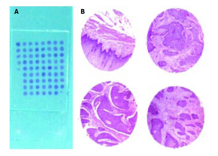
Seventy esophageal squamous carcinoma tissue specimens were selected from Department of Pathology, Shantou University Medical College in 2002. The specimens were fixed in 40 g/L neutrally buffered formaldehyde, embedded in paraffin. Sections of 5 µm stained with hematoxylin and eosin were obtained to confirm the diagnosis and to identify different viable, representative areas of the specimen. From these defined areas core biopsies were taken with a precision instrument. Sixty-eight EC and para-cancerous tissue cores with a diameter of 1.5 mm from each specimen were punched and arrayed in 8 × 9 on a recipient paraffin block. Five-µm sections of the EC tissue array block was cut and placed on adhesive coated slides (cooperated with Cybrdi, Dr. Li Jun).
The expression of Erk1/Erk2 MAPK signaling protein was analyzed using Erk1/Erk2 Mouse derived anti-activated MAP kinase monoclonal antibody (1:400, Sigma), HistostainTM-SP kit and DAB visualization methods according to the manufacture’s instruction (China Beijing Zhongshan Biological Technology CO., LTD.) And the expression of epidermal growth factor receptors were primarily analyzed using phosphor-EGFR (Try845) rabbit polyclonal antibodies and HRP-linked anti-rabbit IgG (Cell Signaling Technology, Inc. #2231, #7074). Four conventional normal esophageal epithelium tissues from autopsy were used as normal tissue controls. The human breast cancer tissue was used as positive control. Negative control was designed using phosphate-buffered saline (PBS) instead of primary antiserum. The detailed immunohistochemical process was carried out according to the manufacture’s instructions.
The positive immunohistochemical staining of Erk1/Erk2 proteins was shown as brown signals in the nuclei; and the immunohistochemical signals of phosphor-EGFR (Tyr845) was in membrane and cytoplasm. The percentage of positive stained cells was evaluated for each tissue sample by counting all cells at 5 high power fields of micrometric rule (5 mm × 5 mm). The cases having positive cancer cells or epithelium accounting for more than 75% of all cancer cells or epithelium on the slide were defined as a score of ++++, 50%-74% were defined as a score of +++, 25-49% were defined as a score of ++, 6-24% were defined as a score of +, 1-5% were defined as a score of ±, less than 1% were defined as a score of -.
Data were stored in a computer data base (FoxPro, version 2.5 b) and analyzed using a computer (Pentium 4) spread sheet (Microsoft Excel 97) and professional statistical computer software (SPSS, version 11.0 and SAS, version 6.08 ). P <= 0.05 was taken as significant. Immunoreactivity was classified as continuous data (undetectable levels or 0% to homogeneous staining or 100%) for all markers.
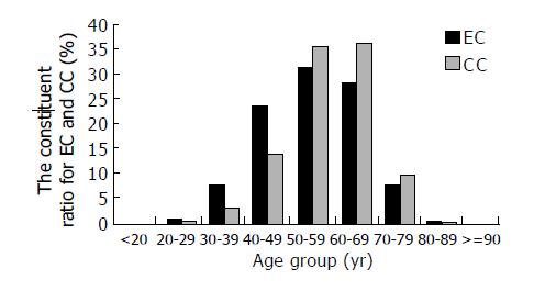
Genders of the patients were recorded in 9 635 cases. The 8 665 EC cases had age records, the youngest and the oldest were 17 years and 91 years respectively, and 3714 CC cases had age records. The male to female ratio and average age of morbidity for EC and CC in Chaoshan region are shown in Table 1. Constituent ratio of EC, CC in every age group is shown in the histogram (Figure 1).
| n | Sex ( male:female) | Age(yr) | F | P | |
| EC | 9 635 | 7 228:2 407(3.0:1) | 54.61 ± 10.73 | 103.24 | < 0.001 |
| CC | 4 167 | 3 442:725(4.75:1) | 58.14 ± 9.45 |
The overwhelming majority of ECs were squamous cell carcinoma (96.4%); whereas CCs were composed mainly of adenocarcinomas (94.5%), and squamous cell carcinoma ranked second (4.4%). The detailed information of pathology is shown in Table 2.
| Site | Gross type | Histological type | |||||||||
| U | M | L | UT | MT | ST | FT | SCA | ACA | UCA | O | |
| EC | 12.7 | 72.0 | 15.3 | 41.5 | 39.6 | 9.6 | 9.4 | 96.4 | 2.7 | 0.6 | 0.3 |
| CC | 4.4 | 94.5 | 0.9 | 0.2 | |||||||
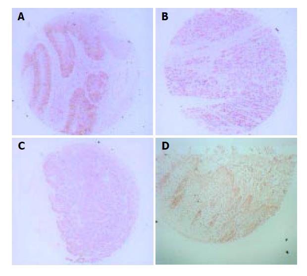
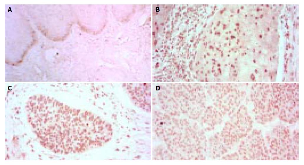
Sections from tissue arrays were kept structure well for pathologic and immunohistochemical research (Figure 2). The expression of phosphor-EGFR(Tyr845) in esophageal squamous carcinoma tissue are relative diversity from ± to ++++, but no distinct differences among esophageal squamous carcinoma tissues according to grading (Figure 3). The expression of activated ERK in esophageal squamous carcinoma tissue and para-cancerous tissue are shown in Table 3 (Figure 4).
| Normal esophagealsquamous cell (n = 5) | Para-canceroustissue (n = 5) | Grade I (n = 19) | Grade II (n = 34) | Grade III (n = 9) | F | P | |
| Activated ERK1/ERK2 | 5.10 ± 1.44 | 76.80 ± 0.14 | 76.80 ± 0.09 | 75.80 ± 0.09 | 69.30 ± 0.14 | 2.60 | 0.04 |
In data from Yangquan city of Shanxi Province (502 cases)[8], the median age of EC patients was 59.17 years, 4.56 years older than that of Chaoshan EC patients. And also data from Linxian of Henan Province (2 601 cases)[9], the proportion of EC patients from 20 to 30 years was 0.35%, from 30 to 40 was 5.19%; whereas both of the two proportions were lower than those from our data (0.70% and 7.70% respectively). In our data from Chaoshan region, the ulcerative type (41.5%) was the most common gross type of EC, which suggests that most EC patients were in the terminal stages when they arrived at hospitals.
Both preceding evidences indicated the average age of EC occurrence was younger in Chaoshan population than those in central plains of China. Synthetically analysis of two reports from Henan Province (1 045 cases and 1 332 cases respectively)[9,10] showed the proportion of CC patients less than 50 years old was 15.56%, which was slightly lower than that from Chaoshan CC data (17.60%). The similar phenomena were seen in other papers[11,12]. It might indicate that the age of incidence of cardiac cancer is also younger in Chaoshan region than in most other areas. Pathological analysis of 1 572 EC patients from Henan Province indicated histological type for the overwhelming majority of EC was squamous cell carcinoma (SCC), and the proportion of SCC (95.1%) was similar to that in our data (96.44%)[13]. In comparison of the constituent ratios of histological types for cardiac carcinoma with data respectively from the Chinese Academy of Medical Science[14], Erlangen-Nurmberg University in Germany[15] and our data, it homoplastically shows that adenocarcinoma accounts for the overwhelming majority of cardiac carcinoma, while the proportions of other histological types are relatively low.
Both data from Yangquan and Linxian[16] also showed the middle third of esophagus was the most commonly affected site analogously, followed by the lower third and then the upper third, similar to our results.
Tissue array technology is applicable for rapid molecular profiling of large numbers of cancers in a single experiment. But a possible limitation of the tissue array technology is that the minute tissue samples acquired from the original tissues may not always be representative of the entire tumor, in light of the intratumor heterogeneity characteristic to most cancers. The comparisons between similarly acquired specimens from different stages of tumor progression placed on the same tissue microarray should be less problematic. If tumor arrays are used to investigate prevalence or prognostic significance of molecular changes, the critical issue is the extent to which minute tissue samples are representative of their donor tumors. The findings of this study suggest that significant results can be obtained on tumor arrays issue cores with a diameter of 1.5 mm. Well and truly, one should consider the tumor tissue array technology as a rapid, high -throughput survey method to pinpoint the biologically most prevalent or clinically most promising genes and molecular markers for detailed studies combined with conventional tissue specimens.
The epidermal growth factor (EGF) peptide induces cellular proliferation through the epidermal growth factor receptor (EGFR), a Mr 170 000 single-pass transmembrane tyrosine kinase, which is believed to play important roles in the control of cell growth and differentiation. The EGFR activates ras and the MAP kinase pathway, ultimately causing phosphorylation of transcription factors such as c-Fos to create AP-1 and ELK-1 that contribute to proliferation. Gene amplification and overexpression of EGFR have been reported in various human tumors, including head and neck/oral cancer[17-22].
Mitogen-activated protein kinase (MAPK) cascades have been shown to play a key role in transduction extracellular signals to cellular responses. Extracellular signal-regulated kinase (ERK) has been the best characterized MAPK and the Raf-MEK-ERK pathway represents one of the best characterized MAPK signaling pathway. The activated ERKs translocate to the nucleus and transactivate transcription factors, changing gene expression to promote growth, differentiation or mitosis[23].
Our primary immunohistochemical study showed that phosphor-EGFR (Tyr845) and activated ERK were expressed in both para-cancerous and esophageal cancerous cells. The intensity of the expression was much higher in cancer than in normal tissue, suggesting that EGFR, Erk1/Erk2 MAPK signaling pathway might play an important role in the regulation of proliferation of esophageal cancer[24,25].
In summary, Both EC and CC are common in males. The average age of occurrence is younger in Chaoshan than in most other regions of China. It is suggested that either genetic factors might play an important role in the pathogenesis of esophageal and cardiac cancers in Chaoshan or Chaoshan residents exposed themselves to some high risk environmental factors. Tissue microarrays technology is applicable for rapid molecular profiling of many tissue samples in a single experiment.
The authors acknowledge the participation of the following members of the undergraduate scientific research group in Shantou University Medical College of China led by Professor Min Su: SM Ying, Y Ni, YS Gao, J Lin, JK Sun, BC Yuan, YF Li, XL Chen, JS X, Y F Chen, who helped us collect the case data of esophageal carcinoma and cardiac cancer. We express our special thanks to Professor Bruce AJ Ponder, Hutchison/MRC Research Centre, MRC Cancer Cell Unit and University of Cambridge, for his invaluable suggestions, Dr. John KL, for his kind assistance in the preparation of the grammatical structure of this paper.
Edited by Zhu LH and Chen WW Proofread by Xu FM
| 1. | Su M, Lu SM, Tian DP, Zhao H, Li XY, Li DR, Zheng ZC. Relationship between ABO blood groups and carcinoma of esophagus and cardia in Chaoshan inhabitants of China. World J Gastroenterol. 2001;7:657-661. [PubMed] |
| 2. | Li K, Su M, Yu P. Mortality trends for malignancies in Nanao county of Guangdong province. Zhongguo Zhongliu. 2001;10:269-270. |
| 3. | Su H, Hu N, Shih J, Hu Y, Wang QH, Chuang EY, Roth MJ, Wang C, Goldstein AM, Ding T. Gene expression analysis of esophageal squamous cell carcinoma reveals consistent molecular profiles related to a family history of upper gastrointestinal cancer. Cancer Res. 2003;63:3872-3876. [PubMed] |
| 4. | Nie Y, Liao J, Zhao X, Song Y, Yang GY, Wang LD, Yang CS. Detection of multiple gene hypermethylation in the development of esophageal squamous cell carcinoma. Carcinogenesis. 2002;23:1713-1720. [RCA] [PubMed] [DOI] [Full Text] [Cited by in Crossref: 60] [Cited by in RCA: 55] [Article Influence: 2.4] [Reference Citation Analysis (0)] |
| 5. | Shinohara M, Aoki T, Sato S, Takagi Y, Osaka Y, Koyanagi Y, Hatooka S, Shinoda M. Cell cycle-regulated factors in esoph-ageal cancer. Dis Esophagus. 2002;15:149-154. [RCA] [DOI] [Full Text] [Cited by in Crossref: 19] [Cited by in RCA: 20] [Article Influence: 0.9] [Reference Citation Analysis (0)] |
| 6. | Gibson MK, Abraham SC, Wu TT, Burtness B, Heitmiller RF, Heath E, Forastiere A. Epidermal growth factor receptor, p53 mutation, and pathological response predict survival in patients with locally advanced esophageal cancer treated with preoperative chemoradiotherapy. Clin Cancer Res. 2003;9:6461-6468. [PubMed] |
| 7. | Arteaga CL. Epidermal growth factor receptor dependence in human tumors: more than just expression. Oncologist. 2002;7 Suppl 4:31-39. [RCA] [PubMed] [DOI] [Full Text] [Cited by in Crossref: 328] [Cited by in RCA: 327] [Article Influence: 14.2] [Reference Citation Analysis (0)] |
| 8. | Li SK, Mao XZ, Jia YT. Clinic analysis of 502 cases with esoph-ageal carcinoma. Zhongliu Yanjiu He Linchuang. 1996;8:106-107. |
| 9. | Wang LD, Gao WJ, Yang WC, Li XF, Li J, Zou JX, Wang DC, Guo RX. Preliminary analysis of the statistics on 3933 cases with esophageal cancer and gastric cardia cancer from the sub-jects in the People's Hospital of Linzhou in 9 years. J Henan Med Univ. 1997;32:9-11. |
| 10. | Collaboration group of gastroscopy in some hospitals of Henan province. Occurrence condition of cardia carcinoma and endo-scope classification of early cardia carcinoma in high and low incidence areas of esophageal carcinoma. J Henan Med Collage. 1987;22:207-210. |
| 11. | Collaboration group of endoscopy in Shanghai. Clinic analysis of 1451 cardia carcinomas diagnosed by gastroscopy. Neijing. 1985;2:32-34. |
| 12. | Hansson LE, Sparén P, Nyrén O. Increasing incidence of carcinoma of the gastric cardia in Sweden from 1970 to 1985. Br J Surg. 1993;80:374-377. [RCA] [PubMed] [DOI] [Full Text] [Cited by in Crossref: 98] [Cited by in RCA: 105] [Article Influence: 3.3] [Reference Citation Analysis (0)] |
| 13. | Xu JY, Zhang JK, Wang RM. Control observation on esophageal carcinoma and esophagitis deteted by endoscopy between high and low incidence areas of esophageal carcinoma. Linchuang Xiaohuabing Zazhi. 1995;7:101-103. |
| 14. | Li L. [Pathologic features of carcinoma of the gastric cardia]. Zhonghua Zhongliu Zazhi. 1984;6:37-40. [PubMed] |
| 15. | Husemann B. Cardia carcinoma considered as a distinct clinical entity. Br J Surg. 1989;76:136-139. [RCA] [PubMed] [DOI] [Full Text] [Cited by in Crossref: 92] [Cited by in RCA: 81] [Article Influence: 2.3] [Reference Citation Analysis (0)] |
| 16. | Liu FS. Pathology of esophageal carcinoma. Zhongliu Fangzhi Yanjiu. 1976;3:234-238. |
| 17. | Eriksen JG, Steiniche T, Askaa J, Alsner J, Overgaard J. The prognostic value of epidermal growth factor receptor is related to tumor differentiation and the overall treatment time of radiotherapy in squamous cell carcinomas of the head and neck. Int J Radiat Oncol Biol Phys. 2004;58:561-566. [RCA] [PubMed] [DOI] [Full Text] [Cited by in Crossref: 89] [Cited by in RCA: 83] [Article Influence: 4.0] [Reference Citation Analysis (0)] |
| 18. | Selvaggi G, Novello S, Torri V, Leonardo E, De Giuli P, Borasio P, Mossetti C, Ardissone F, Lausi P, Scagliotti GV. Epidermal growth factor receptor overexpression correlates with a poor prognosis in completely resected non-small-cell lung cancer. Ann Oncol. 2004;15:28-32. [RCA] [PubMed] [DOI] [Full Text] [Cited by in Crossref: 200] [Cited by in RCA: 214] [Article Influence: 10.2] [Reference Citation Analysis (0)] |
| 19. | Chan KS, Carbajal S, Kiguchi K, Clifford J, Sano S, DiGiovanni J. Epidermal growth factor receptor-mediated activation of Stat3 during multistage skin carcinogenesis. Cancer Research. 2004;64:2382-2389. [RCA] [DOI] [Full Text] [Cited by in Crossref: 134] [Cited by in RCA: 144] [Article Influence: 6.9] [Reference Citation Analysis (0)] |
| 20. | Shimizu M, Suzui M, Deguchi A, Lim JT, Weinstein IB. Ef-fects of acyclic retinoid on growth, cell cycle control, epidermal growth factor receptor signaling, and gene expression in hu-man squamous cell carcinoma cells. Clin Cancer Research. 2004;10:1130-1140. [RCA] [DOI] [Full Text] [Cited by in Crossref: 33] [Cited by in RCA: 34] [Article Influence: 1.6] [Reference Citation Analysis (0)] |
| 21. | Pérez-Soler R. HER1/EGFR targeting: refining the strategy. Oncologist. 2004;9:58-67. [RCA] [PubMed] [DOI] [Full Text] [Cited by in Crossref: 77] [Cited by in RCA: 84] [Article Influence: 4.0] [Reference Citation Analysis (0)] |
| 22. | Dahlberg PS, Ferrin LF, Grindle SM, Nelson CM, Hoang CD, Jacobson B. Gene expression profiles in esophageal adenocarcinoma. Ann Thorac Surg. 2004;77:1008-1015. [RCA] [PubMed] [DOI] [Full Text] [Cited by in Crossref: 33] [Cited by in RCA: 31] [Article Influence: 1.5] [Reference Citation Analysis (0)] |
| 23. | Zhang W, Liu HT. MAPK signal pathways in the regulation of cell proliferation in mammalian cells. Cell Res. 2002;12:9-18. [RCA] [PubMed] [DOI] [Full Text] [Cited by in Crossref: 1583] [Cited by in RCA: 2008] [Article Influence: 87.3] [Reference Citation Analysis (0)] |
| 24. | Nemoto T, Ohashi K, Akashi T, Johnson JD, Hirokawa K. Overexpression of protein tyrosine kinases in human esophageal cancer. Pathobiology. 1997;65:195-203. [RCA] [PubMed] [DOI] [Full Text] [Cited by in Crossref: 98] [Cited by in RCA: 97] [Article Influence: 3.5] [Reference Citation Analysis (0)] |
| 25. | Trisciuoglio D, Iervolino A, Candiloro A, Fibbi G, Fanciulli M, Zangemeister-Wittke U, Zupi G, Del Bufalo D. bcl-2 induction of urokinase plasminogen activator receptor expression in human cancer cells through Sp1 activation: involvement of ERK1/ERK2 activity. J Biol Chem. 2004;279:6737-6745. [RCA] [PubMed] [DOI] [Full Text] [Cited by in Crossref: 53] [Cited by in RCA: 56] [Article Influence: 2.5] [Reference Citation Analysis (0)] |


