Copyright
©The Author(s) 2003.
World J Gastroenterol. Sep 15, 2003; 9(9): 2030-2035
Published online Sep 15, 2003. doi: 10.3748/wjg.v9.i9.2030
Published online Sep 15, 2003. doi: 10.3748/wjg.v9.i9.2030
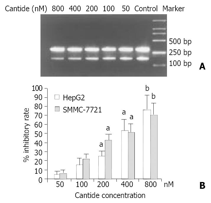
Figure 1 Inhibitory effects of cantide on mRNA level of hTERT.
A: Electrophoresis of PCR products of hTERT gene and β2-microglobulin gene in HepG2 cells treated with cantide. B: Quantitation of inhibitory percentage of hTERT mRNA in treated cells. Each level of PCR product of hTERT gene was quantitated and normalized by the level of β2-microglobulin. Inhibitory rate was calculated by comparing to the control cells. The results were expressed as means ± SD from three inde-pendent experiments. aP < 0.05 vs the cells treated with lipofectin alone, bP < 0.01 vs the cells treated with lipofectin alone.
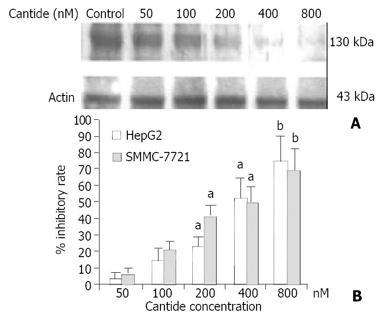
Figure 2 Inhibitory effects of cantide on protein level of hTERT.
A: Western blot analysis of hTERT protein in HepG2 cells treated with cantide. B: Inhibitory percentage of hTERT pro-tein in treated cells compared to the control cells. Each level of hTERT protein was quantitated. Inhibitory rate was calculated by comparing to the control cells. The results were expressed as means ± SD of two independent experiments. aP < 0.05 vs the cells treated with lipofectin alone, bP < 0.01 vs the cells treated with lipofectin alone.
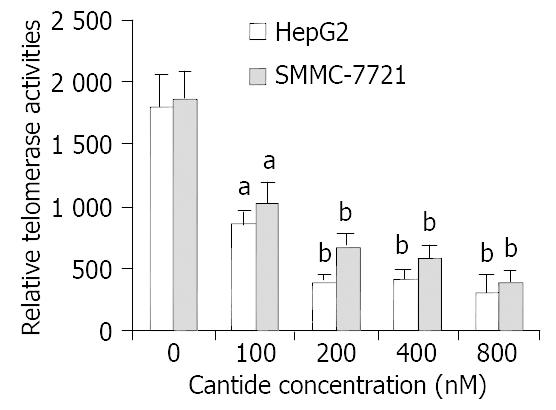
Figure 3 Comparison of telomerase activity in HepG2 and SMMC-7721 cells treated with cantide.
48 h after treatment with cantide, cells extract was tested for telomerase activity by TRAP-ELISA assay. The results were expressed as means ± SD from three independent experiments. aP < 0.05 vs the cells treated with lipofectin alone, bP < 0.01 vs the cells treated with lipofectin alone.
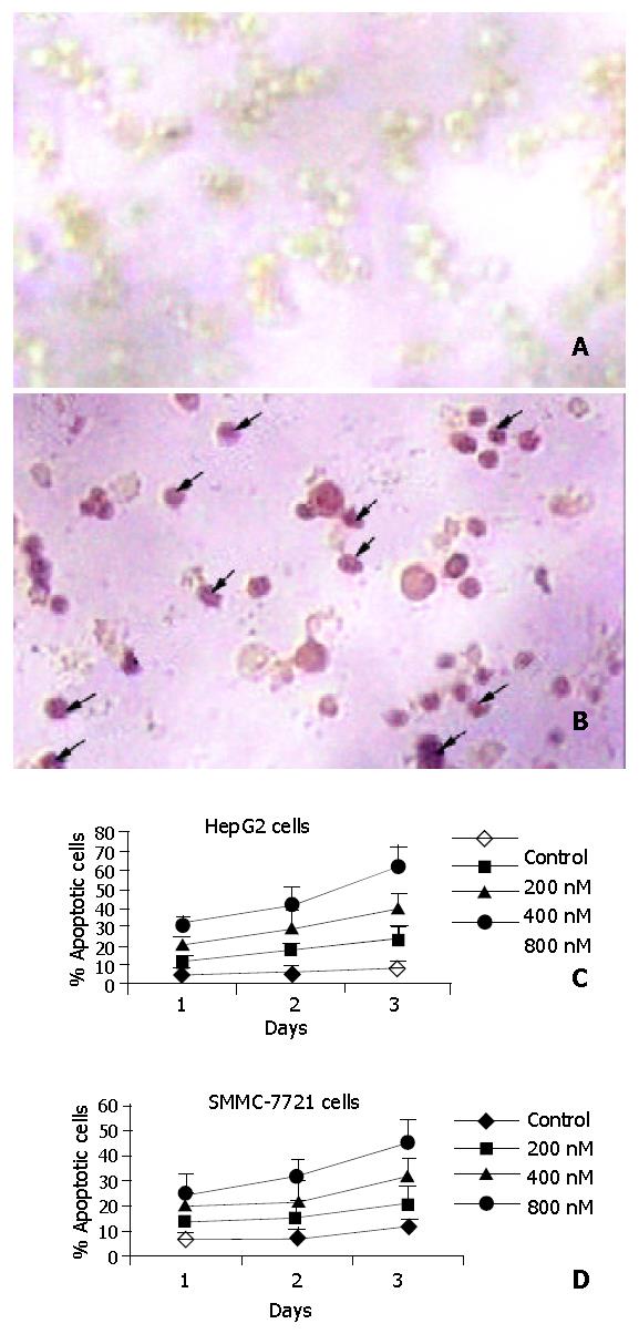
Figure 4 Apoptotic features of tumor cells.
The DeadEnd kit was utilized for the assay. A: staining of the cells treated with (a) lipofectin alone and (b) cantide in combination with lipofectin. The arrows showed the representative cells with dark brown staining, 100 ×. B: the percentage of TUNEL-positive cells treated with cantide. c: HepG2 cells, d: SMMC-7721 cells. The results were expressed as means ± SD from three independent experiments.
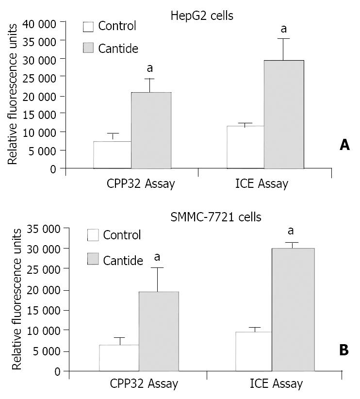
Figure 5 Measurement of CPP32- and ICE-like protease activities in tumor cells treated with cantide.
Two days after treatment with cantide, cell lysates were tested for CPP32-and ICE-like protease activities.The results were expressed as means ± SD from three independent experiments. A: HepG2 cells, B: SMMC-7721 cells. aP < 0.05 vs the cells treated with lipofectin alone.
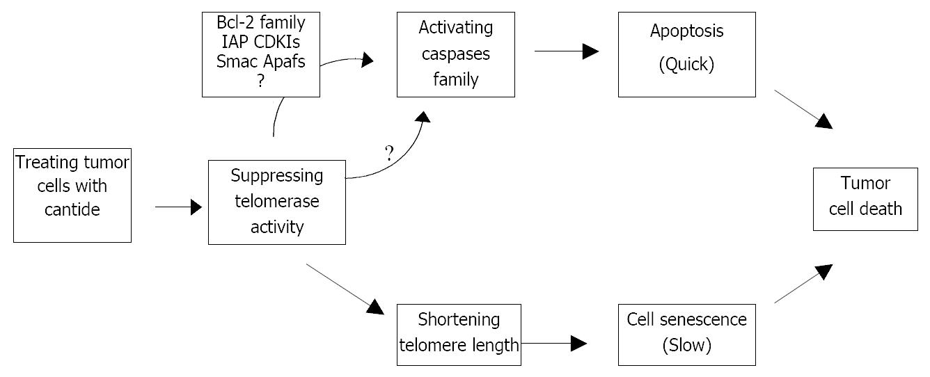
Figure 6 Two possible pathways of antitumor effect of cantide.
IAP: inhibitors of apoptosis, CDKIs: cycin-dependent kinase inhibitors, Apafs: apoptotic protease activating factors.
- Citation: Du QY, Wang XB, Chen XJ, Zheng W, Wang SQ. Antitumor mechanism of antisense cantide targeting human telomerase reverse transcriptase. World J Gastroenterol 2003; 9(9): 2030-2035
- URL: https://www.wjgnet.com/1007-9327/full/v9/i9/2030.htm
- DOI: https://dx.doi.org/10.3748/wjg.v9.i9.2030


