Copyright
©The Author(s) 2003.
World J Gastroenterol. Aug 15, 2003; 9(8): 1795-1798
Published online Aug 15, 2003. doi: 10.3748/wjg.v9.i8.1795
Published online Aug 15, 2003. doi: 10.3748/wjg.v9.i8.1795
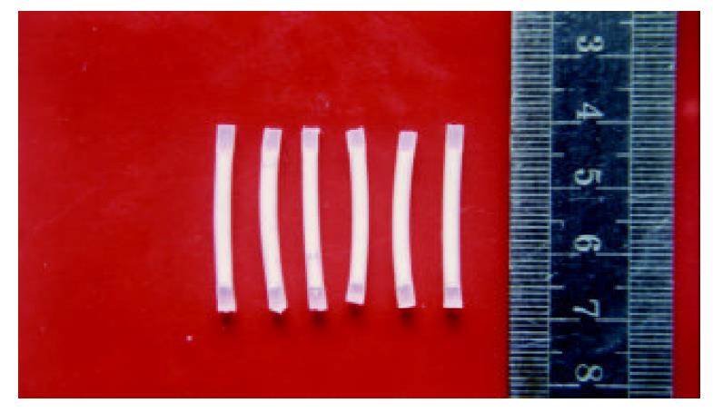
Figure 1 5-FUCI.
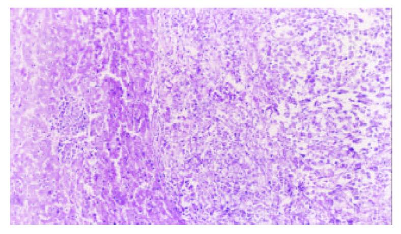
Figure 2 Tumor section (HE×100): tumor cell proliferation was active in group D.
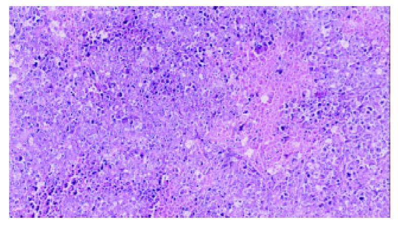
Figure 3 Tumor section (HE×100): a number of focal necrosis observed in group E.
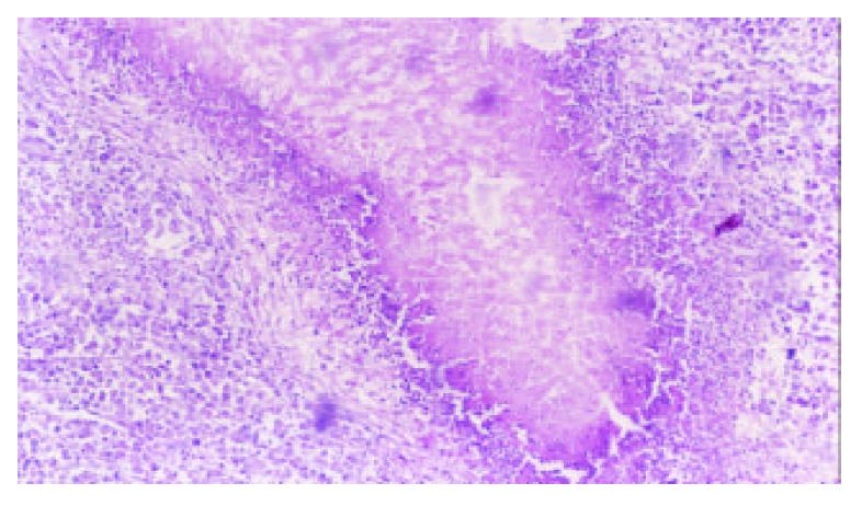
Figure 4 Tumor section (HE×100): the island of necrosis and lymphocytes infiltrated around the necrotic tissue found in group F.
- Citation: He YC, Chen JW, Cao J, Pan DY, Qiao JG. Toxicities and therapeutic effect of 5-fluorouracil controlled release implant on tumor-bearing rats. World J Gastroenterol 2003; 9(8): 1795-1798
- URL: https://www.wjgnet.com/1007-9327/full/v9/i8/1795.htm
- DOI: https://dx.doi.org/10.3748/wjg.v9.i8.1795


