Copyright
©The Author(s) 2003.
World J Gastroenterol. Aug 15, 2003; 9(8): 1772-1776
Published online Aug 15, 2003. doi: 10.3748/wjg.v9.i8.1772
Published online Aug 15, 2003. doi: 10.3748/wjg.v9.i8.1772
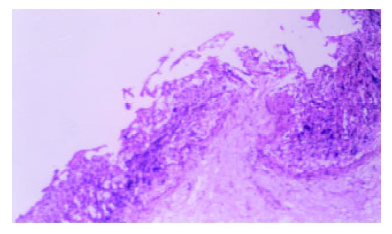
Figure 1 Necrosis appeared as craters in rats after exposed to single WRS for 4 h (HE×200).
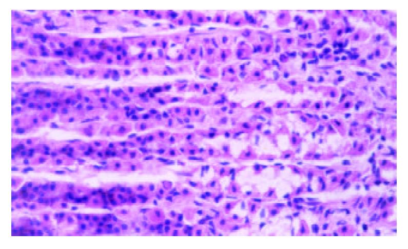
Figure 2 Foveolae and neck region elongated and the mucosa appeared thicker after rats exposed to 4th WRS for 4 h(HE×400).
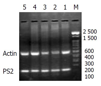
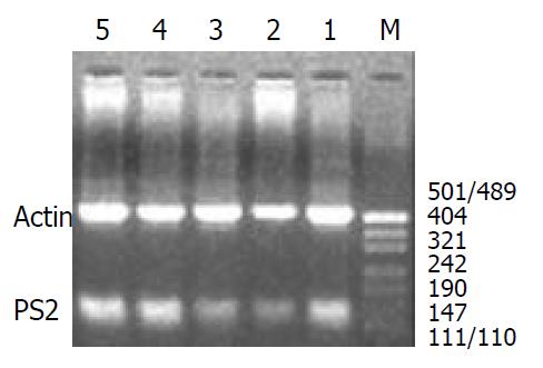
Figure 3 Messenger RNA expression of pS2 mRNA and β-ac-tin in gastric mucosa of rats after single exposure to WRS and in control intact rats (M = PCR size marker, 1 = control group, 2 = group 1, 3 = group 2, 4 = group 3, 5 = group 4).
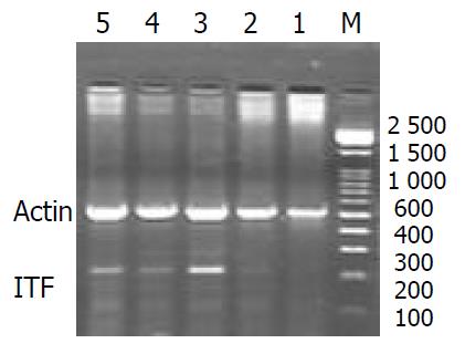
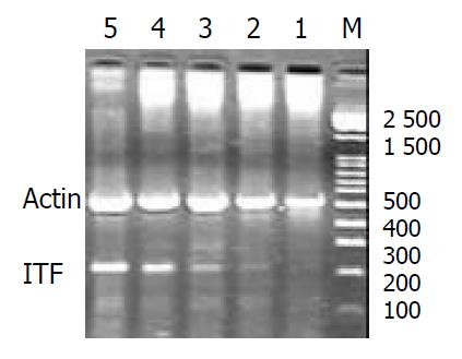
Figure 4 Messenger RNA expression of ITF mRNA and β-actin in gastric mucosa of rats after single exposure to WRS and in control intact rats (M = PCR size marker, 1 = control group, 2 = group 1, 3 = group 2, 4 = group 3, 5 = group 4).
Figure 5 Messenger RNA expression of pS2 mRNA and β-actin in gastric mucosa of rats after repeated exposure to WRS and in control intact rats (M = PCR size marker, 1 = control group, 2 = group I, 3 = group II, 4 = group III, 5 = group IV).
Figure 6 Messenger RNA expression of ITF mRNA and β-actin in gastric mucosa of rats after repeated exposure to WRS and in control intact rats (M = PCR size marker, 1 = control group, 2 = group I, 3 = group II, 4 = group III, 5 = group IV).
- Citation: Nie SN, Qian XM, Wu XH, Yang SY, Tang WJ, Xu BH, Huang F, Lin X, Sun DY, Sun HC, Li ZS. Role of TFF in healing of stress-induced gastric lesions. World J Gastroenterol 2003; 9(8): 1772-1776
- URL: https://www.wjgnet.com/1007-9327/full/v9/i8/1772.htm
- DOI: https://dx.doi.org/10.3748/wjg.v9.i8.1772


