Copyright
©The Author(s) 2003.
World J Gastroenterol. Aug 15, 2003; 9(8): 1702-1706
Published online Aug 15, 2003. doi: 10.3748/wjg.v9.i8.1702
Published online Aug 15, 2003. doi: 10.3748/wjg.v9.i8.1702
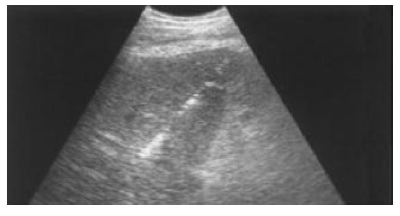
Figure 1 The fine needle was inserted into the right portal vein branch under ultrasound guidance.
After embolization material was injected, tiny spot echoes appeared in the portal vein branch and diffused to the carcinoma area.
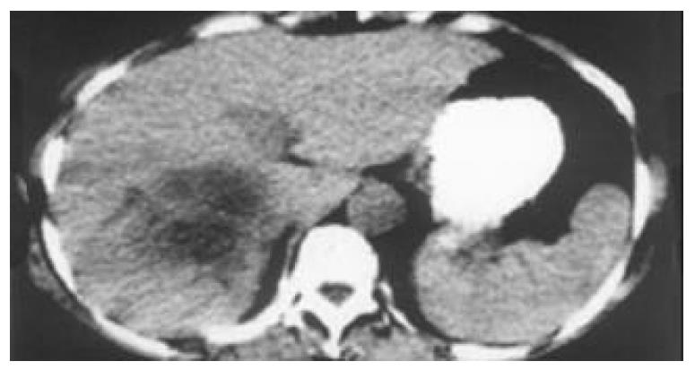
Figure 2 Before POSPVE, CT scan showed a 13.
2 cm×10.8 cm HCC in the right lobe of liver. A right semihepatectomy was scheduled to perform.
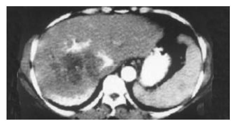
Figure 3 Three weeks after right POSPVE, CT scan showed in-creased volume of left lobe and decreased volume of right lobe.
Iodized oil deposit still could be seen in the right portal vein branch.
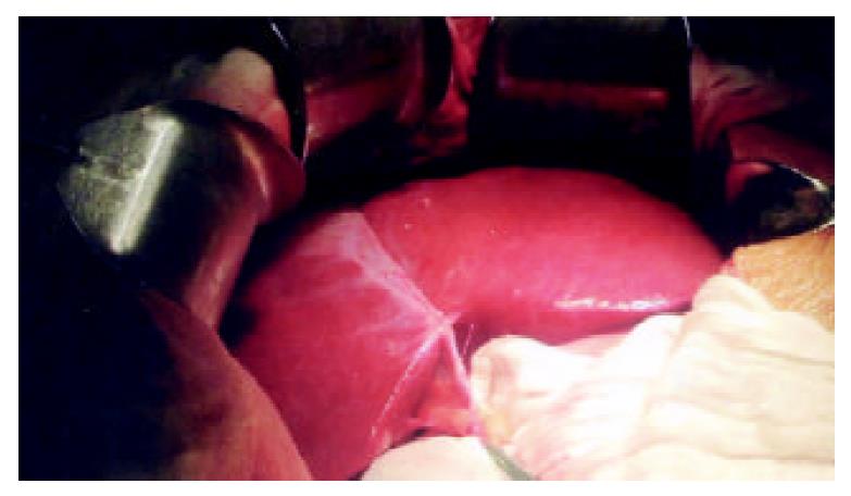
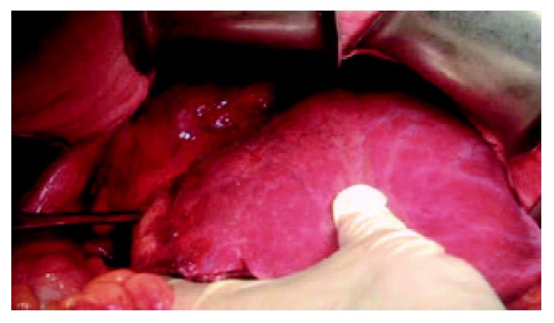
Figure 4 Three weeks after right POSPVE, a right semihepatectomy was performed.
In the operation, significant hypertrophy of left lobe was confirmed.
Figure 5 In the same operation, significant atrophy of right lobe and HCC could be seen.
There was iodized oil deposit in the carcinoma area.
- Citation: Ji W, Li JS, Li LT, Liu WH, Ma KS, Wang XT, He ZP, Dong JH. Role of preoperative selective portal vein embolization in two-step curative hepatectomy for hepatocellular carcinoma. World J Gastroenterol 2003; 9(8): 1702-1706
- URL: https://www.wjgnet.com/1007-9327/full/v9/i8/1702.htm
- DOI: https://dx.doi.org/10.3748/wjg.v9.i8.1702


